 Movie
Movie Controller


+ Open data

- Basic information
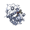
| Entry | Database: PDB / ID: 1pop | ||||||
|---|---|---|---|---|---|---|---|
| Title | X-RAY CRYSTALLOGRAPHIC STRUCTURE OF A PAPAIN-LEUPEPTIN COMPLEX | ||||||
 Components Components |
| ||||||
 Keywords Keywords | HYDROLASE/HYDROLASE INHIBITOR / HYDROLASE-HYDROLASE INHIBITOR COMPLEX | ||||||
| Function / homology |  Function and homology information Function and homology informationpapain / serpin family protein binding / cysteine-type peptidase activity / proteolysis Similarity search - Function | ||||||
| Biological species |  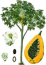 Carica papaya (papaya) Carica papaya (papaya) Streptomyces roseus (bacteria) Streptomyces roseus (bacteria) | ||||||
| Method |  X-RAY DIFFRACTION / Resolution: 2.1 Å X-RAY DIFFRACTION / Resolution: 2.1 Å | ||||||
 Authors Authors | Schroder, E. / Crawford, C. | ||||||
 Citation Citation | Journal: FEBS Lett. / Year: 1993 Title: X-ray crystallographic structure of a papain-leupeptin complex. Authors: Schroder, E. / Phillips, C. / Garman, E. / Harlos, K. / Crawford, C. #1: Journal: J.Mol.Biol. / Year: 1984Title: The Structure of Papain Refined at 1.65 Angstroms Authors: Kamphuis, I.G. / Kalk, K.H. / Swarte, M.B.A. / Drenth, J. | ||||||
| History |
|
- Structure visualization
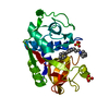
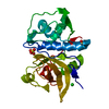
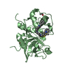
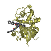
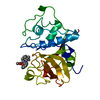
| Structure viewer | Molecule: MolmilJmol/JSmol |
|---|
- Downloads & links
-Download
| PDBx/mmCIF format | 1pop.cif.gz | 74.8 KB | Display | PDBx/mmCIF format |
|---|---|---|---|---|
| PDB format | pdb1pop.ent.gz | 57.1 KB | Display | PDB format |
| PDBx/mmJSON format | 1pop.json.gz | Tree view | PDBx/mmJSON format | |
| Others |  Other downloads Other downloads |
-Validation report
| Arichive directory | https://data.pdbj.org/pub/pdb/validation_reports/po/1popftp://data.pdbj.org/pub/pdb/validation_reports/po/1pop | HTTPS FTP |
|---|
-Related structure data
| Similar structure data |
|---|


-Links
 PDBj
PDBj
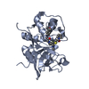
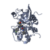
- Assembly
| Deposited unit | 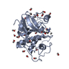
| ||||||||
|---|---|---|---|---|---|---|---|---|---|
| 1 |
| ||||||||
| Unit cell |
| ||||||||
| Atom site foot note | 1: CIS PROLINE - PRO A 152 2: THE LEUPEPTIN MOLECULE, CHAIN *B*, IS COVALENTLY ATTACHED TO CYS 25. |
-Components
| #1: Protein | Mass: 23449.346 Da / Num. of mol.: 1 Source method: isolated from a genetically manipulated source Source: (gene. exp.) Carica papaya (papaya) / References: UniProt: P00784, papain | ||||||||
|---|---|---|---|---|---|---|---|---|---|
| #2: Protein/peptide | 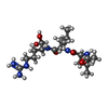  Type: Oligopeptide / Class: Enzyme inhibitor / Mass: 429.578 Da / Num. of mol.: 1 Type: Oligopeptide / Class: Enzyme inhibitor / Mass: 429.578 Da / Num. of mol.: 1Source method: isolated from a genetically manipulated source Source: (gene. exp.) Streptomyces roseus (bacteria) / Strain: MA 839-A1 / References: NOR: NOR00487, LEUPEPTIN | ||||||||
| #3: Chemical | ChemComp-MOH /   Mass: 32.042 Da / Num. of mol.: 24 / Source method: obtained synthetically / Formula: CH4O Mass: 32.042 Da / Num. of mol.: 24 / Source method: obtained synthetically / Formula: CH4O#4: Water | ChemComp-HOH / |  Mass: 18.015 Da / Num. of mol.: 175 / Source method: isolated from a natural source / Formula: H2O Mass: 18.015 Da / Num. of mol.: 175 / Source method: isolated from a natural source / Formula: H2OCompound details | THE LEUPEPTIN IS COVALENTLY | Has protein modification | Y | Sequence details | THERE IS A DISCREPANCY IN THE NORINE AND PDB NUMBERING, AS NORINE COUNTS ACE AND LEU TOGETHER AS ...THERE IS A DISCREPANC | |
-Experimental details
-Experiment
| Experiment | Method: X-RAY DIFFRACTION |
|---|
- Sample preparation
| Crystal | Density Matthews: 2.52 Å3/Da / Density % sol: 51.1 % | ||||||||||||||||||||||||
|---|---|---|---|---|---|---|---|---|---|---|---|---|---|---|---|---|---|---|---|---|---|---|---|---|---|
| Crystal grow | *PLUS Temperature: 4 ℃ / Method: vapor diffusion, sitting drop | ||||||||||||||||||||||||
| Components of the solutions | *PLUS
|
-Data collection
| Radiation | Scattering type: x-ray |
|---|---|
| Radiation wavelength | Relative weight: 1 |
| Reflection | *PLUS Highest resolution: 2.1 Å / Num. obs: 12452 / % possible obs: 88 % / Rmerge(I) obs: 0.094 |
- Processing
| Software |
| ||||||||||||||||||||||||||||||||||||||||||||||||||||||||||||
|---|---|---|---|---|---|---|---|---|---|---|---|---|---|---|---|---|---|---|---|---|---|---|---|---|---|---|---|---|---|---|---|---|---|---|---|---|---|---|---|---|---|---|---|---|---|---|---|---|---|---|---|---|---|---|---|---|---|---|---|---|---|
| Refinement | Highest resolution: 2.1 Å / σ(F): 2 | ||||||||||||||||||||||||||||||||||||||||||||||||||||||||||||
| Refinement step | Cycle: LAST / Highest resolution: 2.1 Å
| ||||||||||||||||||||||||||||||||||||||||||||||||||||||||||||
| Refine LS restraints |
| ||||||||||||||||||||||||||||||||||||||||||||||||||||||||||||
| Refinement | *PLUS Lowest resolution: 20 Å / Rfactor obs: 0.198 | ||||||||||||||||||||||||||||||||||||||||||||||||||||||||||||
| Solvent computation | *PLUS | ||||||||||||||||||||||||||||||||||||||||||||||||||||||||||||
| Displacement parameters | *PLUS |
