 Movie
Movie Controller

[English] 日本語
 Yorodumi
- PDB-1bqi: USE OF PAPAIN AS A MODEL FOR THE STRUCTURE-BASED DESIGN OF CATHEP... -
+ Open data

- Basic information
| Entry | Database: PDB / ID: 1bqi | ||||||
|---|---|---|---|---|---|---|---|
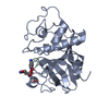
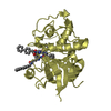
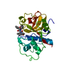
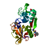
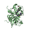
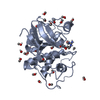
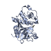
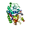
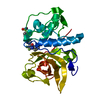
| Title | USE OF PAPAIN AS A MODEL FOR THE STRUCTURE-BASED DESIGN OF CATHEPSIN K INHIBITORS. CRYSTAL STRUCTURES OF TWO PAPAIN INHIBITOR COMPLEXES DEMONSTRATE BINDING TO S'-SUBSITES. | ||||||
 Components Components | PAPAIN | ||||||
 Keywords Keywords | HYDROLASE / SULFHYDRYL PROTEINASE / PAPAIN | ||||||
| Function / homology |  Function and homology information Function and homology informationpapain / serpin family protein binding / cysteine-type peptidase activity / proteolysis Similarity search - Function | ||||||
| Biological species |  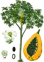 Carica papaya (papaya) Carica papaya (papaya) | ||||||
| Method |  X-RAY DIFFRACTION / MOLECULAR REPLACEMENT / Resolution: 2.5 Å X-RAY DIFFRACTION / MOLECULAR REPLACEMENT / Resolution: 2.5 Å | ||||||
 Authors Authors | Lalonde, J.M. / Zhao, B. / Smith, W.W. / Janson, C.A. / Desjarlais, R.L. / Tomaszek, T.A. / Carr, T.J. / Thompson, S.K. / Yamashita, D.S. / Veber, D.F. / Abdel-Mequid, S.S. | ||||||
 Citation Citation | Journal: J.Med.Chem. / Year: 1998 Title: Use of papain as a model for the structure-based design of cathepsin K inhibitors: crystal structures of two papain-inhibitor complexes demonstrate binding to S'-subsites. Authors: LaLonde, J.M. / Zhao, B. / Smith, W.W. / Janson, C.A. / DesJarlais, R.L. / Tomaszek, T.A. / Carr, T.J. / Thompson, S.K. / Oh, H.J. / Yamashita, D.S. / Veber, D.F. / Abdel-Meguid, S.S. | ||||||
| History |
|
- Structure visualization
| Structure viewer | Molecule: MolmilJmol/JSmol |
|---|
- Downloads & links
-Download
| PDBx/mmCIF format | 1bqi.cif.gz | 52.6 KB | Display | PDBx/mmCIF format |
|---|---|---|---|---|
| PDB format | pdb1bqi.ent.gz | 37.5 KB | Display | PDB format |
| PDBx/mmJSON format | 1bqi.json.gz | Tree view | PDBx/mmJSON format | |
| Others |  Other downloads Other downloads |
-Validation report
| Arichive directory | https://data.pdbj.org/pub/pdb/validation_reports/bq/1bqiftp://data.pdbj.org/pub/pdb/validation_reports/bq/1bqi | HTTPS FTP |
|---|
-Related structure data
| Related structure data |  1bp4C  1pipS S: Starting model for refinement C: citing same article ( |
|---|---|
| Similar structure data |


-Links
 PDBj
PDBj
- Assembly
| Deposited unit | 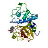
| ||||||||
|---|---|---|---|---|---|---|---|---|---|
| 1 |
| ||||||||
| Unit cell |
|
-Components
| #1: Protein | Mass: 23449.346 Da / Num. of mol.: 1 / Source method: isolated from a natural source Details: METHOXYMETHYLKETONE BOUND NON-COVALENTLY IN S'-SUBSITE Source: (natural) Carica papaya (papaya) / References: UniProt: P00784 |
|---|---|
| #2: Chemical | ChemComp-SBA /   Mass: 408.532 Da / Num. of mol.: 1 / Source method: obtained synthetically / Formula: C22H36N2O5 Mass: 408.532 Da / Num. of mol.: 1 / Source method: obtained synthetically / Formula: C22H36N2O5 |
| #3: Water | ChemComp-HOH /  Mass: 18.015 Da / Num. of mol.: 42 / Source method: isolated from a natural source / Formula: H2O Mass: 18.015 Da / Num. of mol.: 42 / Source method: isolated from a natural source / Formula: H2O |
| Has protein modification | Y |
-Experimental details
-Experiment
| Experiment | Method: X-RAY DIFFRACTION / Number of used crystals: 2 |
|---|
- Sample preparation
| Crystal | Density Matthews: 3.3 Å3/Da / Density % sol: 63.62 % | ||||||||||||||||||||
|---|---|---|---|---|---|---|---|---|---|---|---|---|---|---|---|---|---|---|---|---|---|
| Crystal grow | pH: 8.5 / Details: pH 8.5 | ||||||||||||||||||||
| Crystal grow | *PLUS Method: vapor diffusion, hanging drop | ||||||||||||||||||||
| Components of the solutions | *PLUS
|
-Data collection
| Diffraction | Mean temperature: 287 K |
|---|---|
| Diffraction source | Source: ROTATING ANODE / Type: SIEMENS / Wavelength: 1.5418 |
| Detector | Type: SIEMENS / Detector: AREA DETECTOR / Date: Jun 1, 1995 |
| Radiation | Monochromator: MONOCHROMATOR / Monochromatic (M) / Laue (L): M / Scattering type: x-ray |
| Radiation wavelength | Wavelength: 1.5418 Å / Relative weight: 1 |
| Reflection | Resolution: 2.5→10 Å / Num. obs: 11412 / % possible obs: 91 % / Observed criterion σ(I): 2 / Redundancy: 3.6 % / Rmerge(I) obs: 0.137 / Net I/σ(I): 5.9 |
| Reflection shell | Resolution: 2.54→2.64 Å / Redundancy: 2.9 % / Rmerge(I) obs: 0.37 / Mean I/σ(I) obs: 1.5 / % possible all: 91 |
| Reflection | *PLUS Num. measured all: 41190 |
- Processing
| Software |
| ||||||||||||||||||||||||||||||||||||||||||||||||||||||||||||
|---|---|---|---|---|---|---|---|---|---|---|---|---|---|---|---|---|---|---|---|---|---|---|---|---|---|---|---|---|---|---|---|---|---|---|---|---|---|---|---|---|---|---|---|---|---|---|---|---|---|---|---|---|---|---|---|---|---|---|---|---|---|
| Refinement | Method to determine structure: MOLECULAR REPLACEMENT Starting model: PDB ENTRY 1PIP Resolution: 2.5→10 Å / Data cutoff high absF: 100000 / Data cutoff low absF: 0.1 / Isotropic thermal model: GROUP RESTRAINED / Cross valid method: THROUGHOUT / σ(F): 2 Details: DISORDERED SIDE-CHAINS HAVE THEIR OCCUPANCIES SET TO ZERO IN THE ENTRY, RESIDUES 59, 73, 77, 98, 145
| ||||||||||||||||||||||||||||||||||||||||||||||||||||||||||||
| Displacement parameters | Biso mean: 26.4 Å2 | ||||||||||||||||||||||||||||||||||||||||||||||||||||||||||||
| Refinement step | Cycle: LAST / Resolution: 2.5→10 Å
| ||||||||||||||||||||||||||||||||||||||||||||||||||||||||||||
| Refine LS restraints |
| ||||||||||||||||||||||||||||||||||||||||||||||||||||||||||||
| LS refinement shell | Resolution: 2.5→2.61 Å / Total num. of bins used: 8
| ||||||||||||||||||||||||||||||||||||||||||||||||||||||||||||
| Xplor file |
| ||||||||||||||||||||||||||||||||||||||||||||||||||||||||||||
| Software | *PLUS Name: X-PLOR / Version: 3.1 / Classification: refinement | ||||||||||||||||||||||||||||||||||||||||||||||||||||||||||||
| Refine LS restraints | *PLUS
| ||||||||||||||||||||||||||||||||||||||||||||||||||||||||||||
| LS refinement shell | *PLUS Rfactor Rfree: 0.31 |
