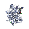
 Movie
Movie Controller


+ Open data

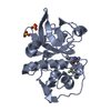
- Basic information
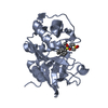
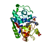
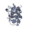
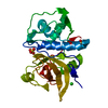
| Entry | Database: PDB / ID: 6ql8 | ||||||
|---|---|---|---|---|---|---|---|
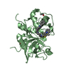
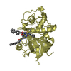
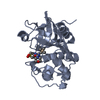
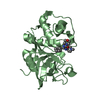
| Title | Cathepsin-K in complex with MIV-711 | ||||||
 Components Components | Cathepsin K | ||||||
 Keywords Keywords | HYDROLASE / inhibitor complex / lysosomal cysteine proteinase | ||||||
| Function / homology |  Function and homology information Function and homology informationcathepsin K / negative regulation of cartilage development / RUNX1 regulates transcription of genes involved in differentiation of keratinocytes / endolysosome lumen / thyroid hormone generation / Trafficking and processing of endosomal TLR / proteoglycan binding / Activation of Matrix Metalloproteinases / Collagen degradation / collagen catabolic process ...cathepsin K / negative regulation of cartilage development / RUNX1 regulates transcription of genes involved in differentiation of keratinocytes / endolysosome lumen / thyroid hormone generation / Trafficking and processing of endosomal TLR / proteoglycan binding / Activation of Matrix Metalloproteinases / Collagen degradation / collagen catabolic process / fibronectin binding / extracellular matrix disassembly / bone resorption / mitophagy / Degradation of the extracellular matrix / collagen binding / cysteine-type peptidase activity / MHC class II antigen presentation / lysosomal lumen / : / lysosome / apical plasma membrane / serine-type endopeptidase activity / external side of plasma membrane / cysteine-type endopeptidase activity / proteolysis / : / extracellular region / nucleoplasm Similarity search - Function | ||||||
| Biological species |  Homo sapiens (human) Homo sapiens (human) | ||||||
| Method |  X-RAY DIFFRACTION / SYNCHROTRON / MOLECULAR REPLACEMENT / molecular replacement / Resolution: 1.8 Å X-RAY DIFFRACTION / SYNCHROTRON / MOLECULAR REPLACEMENT / molecular replacement / Resolution: 1.8 Å | ||||||
 Authors Authors | Derbyshire, D.J. | ||||||
 Citation Citation | Journal: To Be Published Title: Successful development of 3-oxohexahydrofuropyrrole amino acid amides as inhibitors of Cathepsin-K. Authors: Derbyshire, D.J. | ||||||
| History |
|
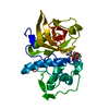
- Structure visualization
| Structure viewer | Molecule: MolmilJmol/JSmol |
|---|
- Downloads & links
-Download
| PDBx/mmCIF format | 6ql8.cif.gz | 104.3 KB | Display | PDBx/mmCIF format |
|---|---|---|---|---|
| PDB format | pdb6ql8.ent.gz | 78.7 KB | Display | PDB format |
| PDBx/mmJSON format | 6ql8.json.gz | Tree view | PDBx/mmJSON format | |
| Others |  Other downloads Other downloads |
-Validation report
| Arichive directory | https://data.pdbj.org/pub/pdb/validation_reports/ql/6ql8ftp://data.pdbj.org/pub/pdb/validation_reports/ql/6ql8 | HTTPS FTP |
|---|
-Related structure data


-Links
 PDBj
PDBj


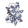
- Assembly
| Deposited unit | 
| ||||||||
|---|---|---|---|---|---|---|---|---|---|
| 1 |
| ||||||||
| Unit cell |
|
-Components
| #1: Protein | Mass: 23737.727 Da / Num. of mol.: 1 Source method: isolated from a genetically manipulated source Source: (gene. exp.) Homo sapiens (human) / Gene: CTSK, CTSO, CTSO2 / Plasmid: pET16b / Production host:  | ||||
|---|---|---|---|---|---|
| #2: Chemical | ChemComp-J5N / ~{  Mass: 569.691 Da / Num. of mol.: 1 / Source method: obtained synthetically / Formula: C29H36FN5O4S / Feature type: SUBJECT OF INVESTIGATION Mass: 569.691 Da / Num. of mol.: 1 / Source method: obtained synthetically / Formula: C29H36FN5O4S / Feature type: SUBJECT OF INVESTIGATION | ||||
| #3: Chemical | ChemComp-NO3 /   Mass: 62.005 Da / Num. of mol.: 6 / Source method: obtained synthetically / Formula: NO3 Mass: 62.005 Da / Num. of mol.: 6 / Source method: obtained synthetically / Formula: NO3#4: Water | ChemComp-HOH / |  Mass: 18.015 Da / Num. of mol.: 260 / Source method: isolated from a natural source / Formula: H2O Mass: 18.015 Da / Num. of mol.: 260 / Source method: isolated from a natural source / Formula: H2OHas protein modification | Y | |
-Experimental details
-Experiment
| Experiment | Method: X-RAY DIFFRACTION / Number of used crystals: 1 |
|---|
- Sample preparation
| Crystal | Density Matthews: 2.87 Å3/Da / Density % sol: 57.2 % |
|---|---|
| Crystal grow | Temperature: 291 K / Method: vapor diffusion, hanging drop / pH: 6 Details: 100mM ADA pH6.0, 500mM ammonium nitrate, 17% PEG 3350 PH range: 6.0-6.2 |
-Data collection
| Diffraction | Mean temperature: 100 K / Serial crystal experiment: N | ||||||||||||||||||||||||
|---|---|---|---|---|---|---|---|---|---|---|---|---|---|---|---|---|---|---|---|---|---|---|---|---|---|
| Diffraction source | Source: SYNCHROTRON / Site: ESRF  / Beamline: ID23-1 / Wavelength: 1.0723 Å / Beamline: ID23-1 / Wavelength: 1.0723 Å | ||||||||||||||||||||||||
| Detector | Type: ADSC QUANTUM 315r / Detector: CCD / Date: Feb 11, 2010 | ||||||||||||||||||||||||
| Radiation | Protocol: SINGLE WAVELENGTH / Monochromatic (M) / Laue (L): M / Scattering type: x-ray | ||||||||||||||||||||||||
| Radiation wavelength | Wavelength: 1.0723 Å / Relative weight: 1 | ||||||||||||||||||||||||
| Reflection | Resolution: 1.8→70.91 Å / Num. obs: 24605 / % possible obs: 98.1 % / Redundancy: 3.9 % / CC1/2: 0.992 / Rmerge(I) obs: 0.117 / Rpim(I) all: 0.067 / Rrim(I) all: 0.136 / Net I/σ(I): 8.5 / Num. measured all: 96143 / Scaling rejects: 8 | ||||||||||||||||||||||||
| Reflection shell | Diffraction-ID: 1
|
-Phasing
| Phasing | Method: molecular replacement |
|---|
- Processing
| Software |
| ||||||||||||||||||||||||||||||||||||||||||||||||||||||||||||
|---|---|---|---|---|---|---|---|---|---|---|---|---|---|---|---|---|---|---|---|---|---|---|---|---|---|---|---|---|---|---|---|---|---|---|---|---|---|---|---|---|---|---|---|---|---|---|---|---|---|---|---|---|---|---|---|---|---|---|---|---|---|
| Refinement | Method to determine structure: MOLECULAR REPLACEMENT / Resolution: 1.8→70.91 Å / Cor.coef. Fo:Fc: 0.965 / Cor.coef. Fo:Fc free: 0.947 / SU B: 4.158 / SU ML: 0.066 / Cross valid method: THROUGHOUT / σ(F): 0 / ESU R: 0.099 / ESU R Free: 0.098 Details: U VALUES : WITH TLS ADDED HYDROGENS HAVE BEEN ADDED IN THE RIDING POSITIONS
| ||||||||||||||||||||||||||||||||||||||||||||||||||||||||||||
| Solvent computation | Ion probe radii: 0.8 Å / Shrinkage radii: 0.8 Å / VDW probe radii: 1.3 Å | ||||||||||||||||||||||||||||||||||||||||||||||||||||||||||||
| Displacement parameters | Biso max: 65.3 Å2 / Biso mean: 17.394 Å2 / Biso min: 8.23 Å2
| ||||||||||||||||||||||||||||||||||||||||||||||||||||||||||||
| Refinement step | Cycle: final / Resolution: 1.8→70.91 Å
| ||||||||||||||||||||||||||||||||||||||||||||||||||||||||||||
| Refine LS restraints |
| ||||||||||||||||||||||||||||||||||||||||||||||||||||||||||||
| LS refinement shell | Resolution: 1.8→1.847 Å / Rfactor Rfree error: 0 / Total num. of bins used: 20
| ||||||||||||||||||||||||||||||||||||||||||||||||||||||||||||
| Refinement TLS params. | Method: refined / Origin x: 27.944 Å / Origin y: 13.763 Å / Origin z: 26.661 Å
|
