 Movie
Movie Controller

[English] 日本語
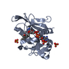
 Yorodumi
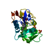
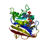
- PDB-1pd8: Analysis of Three Crystal Structure Determinations of a 5-Methyl-... -
+ Open data

- Basic information
| Entry | Database: PDB / ID: 1pd8 | ||||||
|---|---|---|---|---|---|---|---|
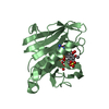
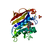
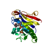
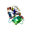
| Title | Analysis of Three Crystal Structure Determinations of a 5-Methyl-6-N-Methylanilino Pyridopyrimidine Antifolate Complex with Human Dihydrofolate Reductase | ||||||
 Components Components | Dihydrofolate reductase | ||||||
 Keywords Keywords | OXIDOREDUCTASE / human dihydrofolate reductase inhibitor complex | ||||||
| Function / homology |  Function and homology information Function and homology informationregulation of removal of superoxide radicals / tetrahydrobiopterin biosynthetic process / Metabolism of folate and pterines / tetrahydrofolate metabolic process / response to methotrexate / sequence-specific mRNA binding / folic acid binding / axon regeneration / dihydrofolate metabolic process / dihydrofolate reductase ...regulation of removal of superoxide radicals / tetrahydrobiopterin biosynthetic process / Metabolism of folate and pterines / tetrahydrofolate metabolic process / response to methotrexate / sequence-specific mRNA binding / folic acid binding / axon regeneration / dihydrofolate metabolic process / dihydrofolate reductase / G1/S-Specific Transcription / dihydrofolate reductase activity / folic acid metabolic process / tetrahydrofolate biosynthetic process / NADPH binding / one-carbon metabolic process / Tetrahydrobiopterin (BH4) synthesis, recycling, salvage and regulation / mRNA regulatory element binding translation repressor activity / NADP binding / molecular adaptor activity / negative regulation of translation / mRNA binding / mitochondrion / cytosol Similarity search - Function | ||||||
| Biological species |  Homo sapiens (human) Homo sapiens (human) | ||||||
| Method |  X-RAY DIFFRACTION / MOLECULAR REPLACEMENT / Resolution: 2.1 Å X-RAY DIFFRACTION / MOLECULAR REPLACEMENT / Resolution: 2.1 Å | ||||||
 Authors Authors | Cody, V. / Luft, J.R. / Pangborn, W. / Gangjee, A. | ||||||
 Citation Citation | Journal: Acta Crystallogr.,Sect.D / Year: 2003 Title: Analysis of three crystal structure determinations of a 5-methyl-6-N-methylanilino pyridopyrimidine antifolate complex with human dihydrofolate reductase. Authors: Cody, V. / Luft, J.R. / Pangborn, W. / Gangjee, A. | ||||||
| History |
|
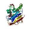
- Structure visualization
| Structure viewer | Molecule: MolmilJmol/JSmol |
|---|
- Downloads & links
-Download
| PDBx/mmCIF format | 1pd8.cif.gz | 56.2 KB | Display | PDBx/mmCIF format |
|---|---|---|---|---|
| PDB format | pdb1pd8.ent.gz | 38.9 KB | Display | PDB format |
| PDBx/mmJSON format | 1pd8.json.gz | Tree view | PDBx/mmJSON format | |
| Others |  Other downloads Other downloads |
-Validation report
| Arichive directory | https://data.pdbj.org/pub/pdb/validation_reports/pd/1pd8ftp://data.pdbj.org/pub/pdb/validation_reports/pd/1pd8 | HTTPS FTP |
|---|
-Related structure data
| Related structure data |  1pd9C  1pdbC  1hfpS C: citing same article ( S: Starting model for refinement |
|---|---|
| Similar structure data |
-Links
 PDBj
PDBj


- Assembly
| Deposited unit | 
| ||||||||
|---|---|---|---|---|---|---|---|---|---|
| 1 |
| ||||||||
| Unit cell |
|
-Components
| #1: Protein | Mass: 21349.525 Da / Num. of mol.: 1 Source method: isolated from a genetically manipulated source Source: (gene. exp.) Homo sapiens (human) / Gene: DHFR / Production host:  |
|---|---|
| #2: Chemical | ChemComp-NDP /   Mass: 745.421 Da / Num. of mol.: 1 / Source method: obtained synthetically / Formula: C21H30N7O17P3 Mass: 745.421 Da / Num. of mol.: 1 / Source method: obtained synthetically / Formula: C21H30N7O17P3 |
| #3: Chemical | ChemComp-CO4 / 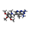  Mass: 384.432 Da / Num. of mol.: 1 / Source method: obtained synthetically / Formula: C19H24N6O3 Mass: 384.432 Da / Num. of mol.: 1 / Source method: obtained synthetically / Formula: C19H24N6O3 |
| #4: Water | ChemComp-HOH /  Mass: 18.015 Da / Num. of mol.: 67 / Source method: isolated from a natural source / Formula: H2O Mass: 18.015 Da / Num. of mol.: 67 / Source method: isolated from a natural source / Formula: H2O |
-Experimental details
-Experiment
| Experiment | Method: X-RAY DIFFRACTION / Number of used crystals: 1 |
|---|
- Sample preparation
| Crystal | Density Matthews: 2.58 Å3/Da / Density % sol: 52.31 % | |||||||||||||||||||||||||||||||||||
|---|---|---|---|---|---|---|---|---|---|---|---|---|---|---|---|---|---|---|---|---|---|---|---|---|---|---|---|---|---|---|---|---|---|---|---|---|
| Crystal grow | Temperature: 293 K / Method: vapor diffusion, hanging drop / pH: 8 Details: Ammonium sulfate, 0.1 M phosphate buffer, pH 8.0, VAPOR DIFFUSION, HANGING DROP, temperature 293K | |||||||||||||||||||||||||||||||||||
| Crystal grow | *PLUS Method: vapor diffusion, hanging drop | |||||||||||||||||||||||||||||||||||
| Components of the solutions | *PLUS
|
-Data collection
| Diffraction | Mean temperature: 298 K |
|---|---|
| Diffraction source | Source: ROTATING ANODE / Type: RIGAKU RU200 / Wavelength: 1.5418 |
| Detector | Type: RIGAKU RAXIS IIC / Detector: IMAGE PLATE / Date: Feb 22, 1994 / Details: mirrors |
| Radiation | Monochromator: graphite / Protocol: SINGLE WAVELENGTH / Monochromatic (M) / Laue (L): M / Scattering type: x-ray |
| Radiation wavelength | Wavelength: 1.5418 Å / Relative weight: 1 |
| Reflection | Resolution: 2.1→8 Å / Num. all: 10192 / Num. obs: 9056 / % possible obs: 82.6 % / Observed criterion σ(F): 2 / Observed criterion σ(I): 1 / Biso Wilson estimate: 25.6 Å2 / Rmerge(I) obs: 0.06 / Rsym value: 0.1 |
| Reflection shell | Resolution: 2.1→2.2 Å / % possible all: 67.4 |
| Reflection | *PLUS Highest resolution: 2 Å / Lowest resolution: 50 Å / Rmerge(I) obs: 0.055 |
| Reflection shell | *PLUS % possible obs: 67.4 % |
- Processing
| Software |
| |||||||||||||||
|---|---|---|---|---|---|---|---|---|---|---|---|---|---|---|---|---|
| Refinement | Method to determine structure: MOLECULAR REPLACEMENT Starting model: PDB entry 1HFP Resolution: 2.1→8 Å / Isotropic thermal model: isotropic / Cross valid method: THROUGHOUT / σ(F): 2 / Stereochemistry target values: Engh & Huber / Details: used weighted full matrix least squares
| |||||||||||||||
| Displacement parameters | Biso mean: 25.6 Å2 | |||||||||||||||
| Refinement step | Cycle: LAST / Resolution: 2.1→8 Å
| |||||||||||||||
| Refine LS restraints |
| |||||||||||||||
| Refine LS restraints | *PLUS
|
