 Movie
Movie Controller

[English] 日本語
 Yorodumi
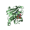
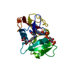
- PDB-3f8z: Human Dihydrofolate Reductase Structural Data with Active Site Mu... -
+ Open data

- Basic information
| Entry | Database: PDB / ID: 3f8z | ||||||
|---|---|---|---|---|---|---|---|
| Title | Human Dihydrofolate Reductase Structural Data with Active Site Mutant Enzyme Complexes | ||||||
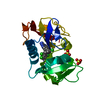
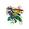
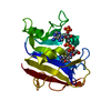
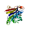
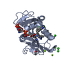
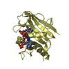
 Components Components | Dihydrofolate reductase | ||||||
 Keywords Keywords | OXIDOREDUCTASE / active site mutants ligand complex human dihydrofolate reductase / NADP / One-carbon metabolism | ||||||
| Function / homology |  Function and homology information Function and homology informationregulation of removal of superoxide radicals / tetrahydrobiopterin biosynthetic process / Metabolism of folate and pterines / tetrahydrofolate metabolic process / response to methotrexate / sequence-specific mRNA binding / folic acid binding / axon regeneration / dihydrofolate metabolic process / dihydrofolate reductase ...regulation of removal of superoxide radicals / tetrahydrobiopterin biosynthetic process / Metabolism of folate and pterines / tetrahydrofolate metabolic process / response to methotrexate / sequence-specific mRNA binding / folic acid binding / axon regeneration / dihydrofolate metabolic process / dihydrofolate reductase / G1/S-Specific Transcription / dihydrofolate reductase activity / folic acid metabolic process / tetrahydrofolate biosynthetic process / NADPH binding / 'de novo' pyrimidine nucleobase biosynthetic process / one-carbon metabolic process / Tetrahydrobiopterin (BH4) synthesis, recycling, salvage and regulation / mRNA regulatory element binding translation repressor activity / replication fork / NADP binding / DNA replication / negative regulation of translation / mRNA binding / mitochondrion / nucleus / cytosol Similarity search - Function | ||||||
| Biological species |  Homo sapiens (human) Homo sapiens (human) | ||||||
| Method |  X-RAY DIFFRACTION / MOLECULAR REPLACEMENT / Resolution: 2.01 Å X-RAY DIFFRACTION / MOLECULAR REPLACEMENT / Resolution: 2.01 Å | ||||||
 Authors Authors | Cody, V. / Pace, J. / Makin, J. / Piraino, J. / Queener, S.F. / Rosowsky, A. | ||||||
 Citation Citation | Journal: Biochemistry / Year: 2009 Title: Correlations of Inhibitor Kinetics for Pneumocystis jirovecii and Human Dihydrofolate Reductase with Structural Data for Human Active Site Mutant Enzyme Complexes. Authors: Cody, V. / Pace, J. / Makin, J. / Piraino, J. / Queener, S.F. / Rosowsky, A. | ||||||
| History |
|
- Structure visualization
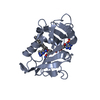
| Structure viewer | Molecule: MolmilJmol/JSmol |
|---|
- Downloads & links
-Download
| PDBx/mmCIF format | 3f8z.cif.gz | 63.4 KB | Display | PDBx/mmCIF format |
|---|---|---|---|---|
| PDB format | pdb3f8z.ent.gz | 44.2 KB | Display | PDB format |
| PDBx/mmJSON format | 3f8z.json.gz | Tree view | PDBx/mmJSON format | |
| Others |  Other downloads Other downloads |
-Validation report
| Arichive directory | https://data.pdbj.org/pub/pdb/validation_reports/f8/3f8zftp://data.pdbj.org/pub/pdb/validation_reports/f8/3f8z | HTTPS FTP |
|---|
-Related structure data
| Related structure data |  3f8yC  3f91C  3fs6C 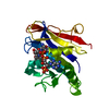 1u72S C: citing same article ( S: Starting model for refinement |
|---|---|
| Similar structure data |


-Links
 PDBj
PDBj


- Assembly
| Deposited unit | 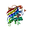
| ||||||||
|---|---|---|---|---|---|---|---|---|---|
| 1 |
| ||||||||
| Unit cell |
|
-Components
| #1: Protein | Mass: 21412.646 Da / Num. of mol.: 1 / Mutation: Q35S, N64S Source method: isolated from a genetically manipulated source Source: (gene. exp.) Homo sapiens (human) / Gene: DHFR, DHFRP1 / Production host:  | ||
|---|---|---|---|
| #2: Chemical | ChemComp-DH1 /   Mass: 346.381 Da / Num. of mol.: 1 / Source method: obtained synthetically / Formula: C17H22N4O4 Mass: 346.381 Da / Num. of mol.: 1 / Source method: obtained synthetically / Formula: C17H22N4O4 | ||
| #3: Chemical | ChemComp-NDP /   Mass: 745.421 Da / Num. of mol.: 1 / Source method: obtained synthetically / Formula: C21H30N7O17P3 Mass: 745.421 Da / Num. of mol.: 1 / Source method: obtained synthetically / Formula: C21H30N7O17P3 | ||
| #4: Chemical | ChemComp-SO4 /   Mass: 96.063 Da / Num. of mol.: 5 / Source method: obtained synthetically / Formula: SO4 Mass: 96.063 Da / Num. of mol.: 5 / Source method: obtained synthetically / Formula: SO4#5: Water | ChemComp-HOH / |  Mass: 18.015 Da / Num. of mol.: 276 / Source method: isolated from a natural source / Formula: H2O Mass: 18.015 Da / Num. of mol.: 276 / Source method: isolated from a natural source / Formula: H2O |
-Experimental details
-Experiment
| Experiment | Method: X-RAY DIFFRACTION / Number of used crystals: 1 |
|---|
- Sample preparation
| Crystal | Density Matthews: 2.51 Å3/Da / Density % sol: 51.02 % |
|---|---|
| Crystal grow | Temperature: 287 K / Method: vapor diffusion, hanging drop / pH: 6.9 Details: 100 mM potassium phosphate, pH 6.9, 60% ammonium sulfate, 3% ethanol, VAPOR DIFFUSION, HANGING DROP, temperature 287K |
-Data collection
| Diffraction | Mean temperature: 298 K |
|---|---|
| Diffraction source | Source: ROTATING ANODE / Type: RIGAKU RU300 / Wavelength: 1.5418 Å |
| Detector | Type: RIGAKU RAXIS IV / Detector: IMAGE PLATE / Date: Mar 10, 2008 / Details: mirrors |
| Radiation | Monochromator: graphite / Protocol: SINGLE WAVELENGTH / Monochromatic (M) / Laue (L): M / Scattering type: x-ray |
| Radiation wavelength | Wavelength: 1.5418 Å / Relative weight: 1 |
| Reflection | Resolution: 1.68→26.25 Å / Num. obs: 17593 / % possible obs: 74.8 % / Observed criterion σ(F): 2 / Observed criterion σ(I): 1 / Redundancy: 3.9 % / Biso Wilson estimate: 32.56 Å2 / Rmerge(I) obs: 0.071 / Rsym value: 0.039 / Net I/σ(I): 16.3 |
| Reflection shell | Resolution: 1.68→1.77 Å / Redundancy: 1.7 % / Rmerge(I) obs: 0.297 / Mean I/σ(I) obs: 1.3 / Num. unique all: 590 / Rsym value: 0.261 / % possible all: 17 |
- Processing
| Software |
| ||||||||||||||||||||||||||||||||||||||||||||||||||||||||||||||||||||||||||||||||||||||||||
|---|---|---|---|---|---|---|---|---|---|---|---|---|---|---|---|---|---|---|---|---|---|---|---|---|---|---|---|---|---|---|---|---|---|---|---|---|---|---|---|---|---|---|---|---|---|---|---|---|---|---|---|---|---|---|---|---|---|---|---|---|---|---|---|---|---|---|---|---|---|---|---|---|---|---|---|---|---|---|---|---|---|---|---|---|---|---|---|---|---|---|---|
| Refinement | Method to determine structure: MOLECULAR REPLACEMENT Starting model: 1u72 Resolution: 2.01→26.66 Å / Cor.coef. Fo:Fc: 0.954 / Cor.coef. Fo:Fc free: 0.93 / SU B: 3.743 / SU ML: 0.106 / Cross valid method: THROUGHOUT / σ(F): 2 / σ(I): 1 / ESU R: 0.199 / ESU R Free: 0.174 / Stereochemistry target values: MAXIMUM LIKELIHOOD / Details: HYDROGENS HAVE BEEN ADDED IN THE RIDING POSITIONS
| ||||||||||||||||||||||||||||||||||||||||||||||||||||||||||||||||||||||||||||||||||||||||||
| Solvent computation | Ion probe radii: 0.8 Å / Shrinkage radii: 0.8 Å / VDW probe radii: 1.2 Å / Solvent model: MASK | ||||||||||||||||||||||||||||||||||||||||||||||||||||||||||||||||||||||||||||||||||||||||||
| Displacement parameters | Biso mean: 19.993 Å2
| ||||||||||||||||||||||||||||||||||||||||||||||||||||||||||||||||||||||||||||||||||||||||||
| Refine analyze | Luzzati coordinate error obs: 0.186 Å | ||||||||||||||||||||||||||||||||||||||||||||||||||||||||||||||||||||||||||||||||||||||||||
| Refinement step | Cycle: LAST / Resolution: 2.01→26.66 Å
| ||||||||||||||||||||||||||||||||||||||||||||||||||||||||||||||||||||||||||||||||||||||||||
| Refine LS restraints |
| ||||||||||||||||||||||||||||||||||||||||||||||||||||||||||||||||||||||||||||||||||||||||||
| LS refinement shell | Resolution: 2.01→2.062 Å / Total num. of bins used: 20
|
