 Movie
Movie Controller


+ Open data

- Basic information
| Entry | Database: PDB / ID: 1pb0 | ||||||
|---|---|---|---|---|---|---|---|
| Title | YCDX PROTEIN IN AUTOINHIBITED STATE | ||||||
 Components Components | Hypothetical protein ycdX | ||||||
 Keywords Keywords | STRUCTURAL GENOMICS / UNKNOWN FUNCTION / beta-alpha-barrel / trinuclear zinc / autoinhibition | ||||||
| Function / homology |  Function and homology information Function and homology informationphosphoric ester hydrolase activity / Hydrolases; Acting on ester bonds; Phosphoric-monoester hydrolases / bacterial-type flagellum-dependent swarming motility / phosphatase activity / zinc ion binding / identical protein binding / cytosol Similarity search - Function | ||||||
| Biological species |  | ||||||
| Method |  X-RAY DIFFRACTION / SYNCHROTRON / MOLECULAR REPLACEMENT / Resolution: 1.95 Å X-RAY DIFFRACTION / SYNCHROTRON / MOLECULAR REPLACEMENT / Resolution: 1.95 Å | ||||||
 Authors Authors | Teplyakov, A. / Obmolova, G. / Khil, P.P. / Camerini-Otero, R.D. / Gilliland, G.L. | ||||||
 Citation Citation | Journal: To be Published Title: Autoregulation of YcdX protein Authors: Teplyakov, A. / Obmolova, G. / Khil, P.P. / Camerini-Otero, R.D. / Gilliland, G.L. #1: Journal: Proteins / Year: 2003Title: Crystal structure of the Escherichia coli YcdX protein reveals a trinuclear zinc active site. Authors: Teplyakov, A. / Obmolova, G. / Khil, P.P. / Howard, A.J. / Camerini-Otero, R.D. / Gilliland, G.L. | ||||||
| History |
|
- Structure visualization
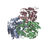
| Structure viewer | Molecule: MolmilJmol/JSmol |
|---|
- Downloads & links
-Download
| PDBx/mmCIF format | 1pb0.cif.gz | 165.2 KB | Display | PDBx/mmCIF format |
|---|---|---|---|---|
| PDB format | pdb1pb0.ent.gz | 128.9 KB | Display | PDB format |
| PDBx/mmJSON format | 1pb0.json.gz | Tree view | PDBx/mmJSON format | |
| Others |  Other downloads Other downloads |
-Validation report
| Arichive directory | https://data.pdbj.org/pub/pdb/validation_reports/pb/1pb0ftp://data.pdbj.org/pub/pdb/validation_reports/pb/1pb0 | HTTPS FTP |
|---|
-Related structure data
| Related structure data |  1m65S S: Starting model for refinement |
|---|---|
| Similar structure data |

-Links
 PDBj
PDBj
- Assembly
| Deposited unit | 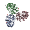
| ||||||||
|---|---|---|---|---|---|---|---|---|---|
| 1 |
| ||||||||
| Unit cell |
| ||||||||
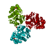
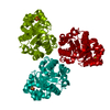
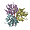
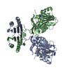
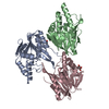
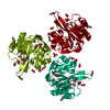
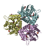
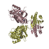
| Details | The asymmetric unit contains the entire trimer that corresponds to the oligomeric state of the protein in solution |
-Components
| #1: Protein | Mass: 26925.529 Da / Num. of mol.: 3 Source method: isolated from a genetically manipulated source Source: (gene. exp.) #2: Chemical | ChemComp-ZN /   Mass: 65.409 Da / Num. of mol.: 9 / Source method: obtained synthetically / Formula: Zn Mass: 65.409 Da / Num. of mol.: 9 / Source method: obtained synthetically / Formula: Zn#3: Chemical | ChemComp-SO4 /   Mass: 96.063 Da / Num. of mol.: 4 / Source method: obtained synthetically / Formula: SO4 Mass: 96.063 Da / Num. of mol.: 4 / Source method: obtained synthetically / Formula: SO4#4: Chemical | ChemComp-FMT /   Mass: 46.025 Da / Num. of mol.: 6 / Source method: obtained synthetically / Formula: CH2O2 Mass: 46.025 Da / Num. of mol.: 6 / Source method: obtained synthetically / Formula: CH2O2#5: Water | ChemComp-HOH / |  Mass: 18.015 Da / Num. of mol.: 699 / Source method: isolated from a natural source / Formula: H2O Mass: 18.015 Da / Num. of mol.: 699 / Source method: isolated from a natural source / Formula: H2O |
|---|
-Experimental details
-Experiment
| Experiment | Method: X-RAY DIFFRACTION / Number of used crystals: 1 |
|---|
- Sample preparation
| Crystal | Density Matthews: 2.57 Å3/Da / Density % sol: 52.15 % |
|---|---|
| Crystal grow | Temperature: 295 K / Method: vapor diffusion, hanging drop / pH: 8.5 Details: 0.1 M Tris, 60% AS, 3% MPD, pH 8.5, VAPOR DIFFUSION, HANGING DROP, temperature 295K |
-Data collection
| Diffraction | Mean temperature: 100 K |
|---|---|
| Diffraction source | Source: SYNCHROTRON / Site: APS  / Beamline: 17-BM / Wavelength: 1 Å / Beamline: 17-BM / Wavelength: 1 Å |
| Detector | Type: MARRESEARCH / Detector: CCD / Date: Mar 15, 2003 / Details: mirror |
| Radiation | Monochromator: graphite / Protocol: SINGLE WAVELENGTH / Monochromatic (M) / Laue (L): M / Scattering type: x-ray |
| Radiation wavelength | Wavelength: 1 Å / Relative weight: 1 |
| Reflection | Resolution: 1.95→30 Å / Num. all: 59787 / Num. obs: 59787 / % possible obs: 99.8 % / Observed criterion σ(F): 0 / Observed criterion σ(I): -3 / Redundancy: 4.7 % / Biso Wilson estimate: 17.5 Å2 / Rmerge(I) obs: 0.081 / Net I/σ(I): 16.2 |
| Reflection shell | Resolution: 1.95→2 Å / Redundancy: 4.7 % / Rmerge(I) obs: 0.212 / Mean I/σ(I) obs: 3.3 / Num. unique all: 7470 / % possible all: 98.6 |
- Processing
| Software |
| ||||||||||||||||||||||||||||||||||||||||||||||||||||||||||||||||||||||
|---|---|---|---|---|---|---|---|---|---|---|---|---|---|---|---|---|---|---|---|---|---|---|---|---|---|---|---|---|---|---|---|---|---|---|---|---|---|---|---|---|---|---|---|---|---|---|---|---|---|---|---|---|---|---|---|---|---|---|---|---|---|---|---|---|---|---|---|---|---|---|---|
| Refinement | Method to determine structure: MOLECULAR REPLACEMENT Starting model: 1M65 Resolution: 1.95→10 Å / Cor.coef. Fo:Fc: 0.949 / SU B: 1.628 / SU ML: 0.05 / σ(F): 0 / ESU R: 0.135 / Stereochemistry target values: Engh & Huber
| ||||||||||||||||||||||||||||||||||||||||||||||||||||||||||||||||||||||
| Solvent computation | Ion probe radii: 0.8 Å / Shrinkage radii: 0.8 Å / VDW probe radii: 1.4 Å / Solvent model: BABINET MODEL WITH MASK | ||||||||||||||||||||||||||||||||||||||||||||||||||||||||||||||||||||||
| Displacement parameters | Biso mean: 24.717 Å2
| ||||||||||||||||||||||||||||||||||||||||||||||||||||||||||||||||||||||
| Refine analyze | Luzzati coordinate error obs: 0.135 Å | ||||||||||||||||||||||||||||||||||||||||||||||||||||||||||||||||||||||
| Refinement step | Cycle: LAST / Resolution: 1.95→10 Å
| ||||||||||||||||||||||||||||||||||||||||||||||||||||||||||||||||||||||
| Refine LS restraints |
| ||||||||||||||||||||||||||||||||||||||||||||||||||||||||||||||||||||||
| LS refinement shell | Resolution: 1.95→2 Å / Total num. of bins used: 20 /
|
