retinal rod cell apoptotic process / PDE3B signalling / cellular response to high light intensity / Inhibition of TSC complex formation by PKB / AKT-mediated inactivation of FOXO1A / Negative regulation of the PI3K/AKT network / negative regulation of long-chain fatty acid import across plasma membrane / Activation of AKT2 / AKT phosphorylates targets in the nucleus / RUNX2 regulates genes involved in cell migration ...retinal rod cell apoptotic process / PDE3B signalling / cellular response to high light intensity / Inhibition of TSC complex formation by PKB / AKT-mediated inactivation of FOXO1A / Negative regulation of the PI3K/AKT network / negative regulation of long-chain fatty acid import across plasma membrane / Activation of AKT2 / AKT phosphorylates targets in the nucleus / RUNX2 regulates genes involved in cell migration / : / positive regulation of fatty acid beta-oxidation / mammary gland epithelial cell differentiation / RAB GEFs exchange GTP for GDP on RABs / positive regulation of glucose metabolic process / peripheral nervous system myelin maintenance / positive regulation of cell motility / glycogen biosynthetic process / AKT phosphorylates targets in the cytosol / Regulation of TP53 Activity through Association with Co-factors / CTLA4 inhibitory signaling / fat cell differentiation / Constitutive Signaling by AKT1 E17K in Cancer / CD28 dependent PI3K/Akt signaling / Regulation of localization of FOXO transcription factors / positive regulation of glycogen biosynthetic process / Estrogen-dependent nuclear events downstream of ESR-membrane signaling / Activation of BAD and translocation to mitochondria / positive regulation of protein targeting to membrane / SARS-CoV-2 targets host intracellular signalling and regulatory pathways / Cyclin E associated events during G1/S transition / Cyclin A:Cdk2-associated events at S phase entry / regulation of cell migration / Regulation of TP53 Activity through Acetylation / FLT3 Signaling / Downregulation of ERBB2:ERBB3 signaling / VEGFR2 mediated vascular permeability / molecular function activator activity / protein localization to plasma membrane / Translocation of SLC2A4 (GLUT4) to the plasma membrane / Deactivation of the beta-catenin transactivating complex / positive regulation of glucose import / TP53 Regulates Metabolic Genes / protein modification process / ruffle membrane / Regulation of PTEN stability and activity / G beta:gamma signalling through PI3Kgamma / cellular response to insulin stimulus / glucose metabolic process / KEAP1-NFE2L2 pathway / Regulation of TP53 Degradation / insulin receptor signaling pathway / PIP3 activates AKT signaling / regulation of translation / cell cortex / early endosome / non-specific serine/threonine protein kinase / regulation of cell cycle / intracellular signal transduction / positive regulation of cell migration / phosphorylation / intracellular membrane-bounded organelle / protein serine kinase activity / protein serine/threonine kinase activity / signal transduction / nucleoplasm / ATP binding / nucleus / metal ion binding / plasma membrane / cytosol 類似検索 - 分子機能
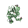
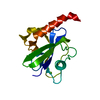
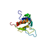
Protein Kinase B beta, catalytic domain / Protein Kinase B, pleckstrin homology domain / Protein kinase, C-terminal / Protein kinase C terminal domain / Pleckstrin-homology domain (PH domain)/Phosphotyrosine-binding domain (PTB) / PH-domain like / Extension to Ser/Thr-type protein kinases / AGC-kinase, C-terminal / AGC-kinase C-terminal domain profile. / PH domain ...Protein Kinase B beta, catalytic domain / Protein Kinase B, pleckstrin homology domain / Protein kinase, C-terminal / Protein kinase C terminal domain / Pleckstrin-homology domain (PH domain)/Phosphotyrosine-binding domain (PTB) / PH-domain like / Extension to Ser/Thr-type protein kinases / AGC-kinase, C-terminal / AGC-kinase C-terminal domain profile. / PH domain / PH domain profile. / Pleckstrin homology domain. / Pleckstrin homology domain / PH-like domain superfamily / Roll / Serine/threonine-protein kinase, active site / Serine/Threonine protein kinases active-site signature. / Protein kinase domain / Serine/Threonine protein kinases, catalytic domain / Protein kinase, ATP binding site / Protein kinases ATP-binding region signature. / Protein kinase domain profile. / Protein kinase domain / Protein kinase-like domain superfamily / Mainly Beta 類似検索 - ドメイン・相同性
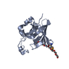
手法: distance geometry, simulated annealing / ソフトェア番号: 1 詳細: The structures are based on a total of 1229 restraints, 1034 are NOE-derived distance constraints, 127 dihedral angle restraints, 68 distance restraints from hydrogen bonds. There are no ...詳細: The structures are based on a total of 1229 restraints, 1034 are NOE-derived distance constraints, 127 dihedral angle restraints, 68 distance restraints from hydrogen bonds. There are no constraints for the two peptidic segments: E59-Q61 and R76-T87.
代表構造
選択基準: lowest energy
NMRアンサンブル
コンフォーマー選択の基準: structures with the lowest energy 計算したコンフォーマーの数: 80 / 登録したコンフォーマーの数: 20
 ムービー
ムービー コントローラー


 データを開く
 基本情報
基本情報 要素
要素 キーワード
キーワード 機能・相同性情報
 Homo sapiens (ヒト)
 データ登録者
 引用
引用 構造の表示
構造の表示 ダウンロードとリンク
 その他のダウンロード
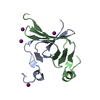


 PDBj
PDBj


 集合体
集合体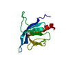

 試料調製
試料調製 解析
解析 XPLOR
XPLOR