 Movie
Movie Controller

[English] 日本語
 Yorodumi
- PDB-4ic9: Crystal structure of the full-length matrix subunit (p15) of the ... -
+ Open data

- Basic information
| Entry | Database: PDB / ID: 4ic9 | ||||||
|---|---|---|---|---|---|---|---|
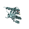
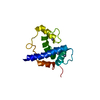
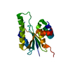
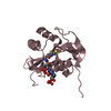
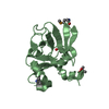
| Title | Crystal structure of the full-length matrix subunit (p15) of the Feline Immunodeficiency Virus (FIV) Gag polyprotein | ||||||
 Components Components | Matrix protein p15 | ||||||
 Keywords Keywords | VIRAL PROTEIN / FIV / p15 / Retroviral matrix protein | ||||||
| Function / homology |  Function and homology information Function and homology informationviral budding via host ESCRT complex / viral nucleocapsid / structural constituent of virion / nucleic acid binding / zinc ion binding Similarity search - Function | ||||||
| Biological species |  Feline immunodeficiency virus Feline immunodeficiency virus | ||||||
| Method |  X-RAY DIFFRACTION / SYNCHROTRON / MOLECULAR REPLACEMENT / Resolution: 2 Å X-RAY DIFFRACTION / SYNCHROTRON / MOLECULAR REPLACEMENT / Resolution: 2 Å | ||||||
 Authors Authors | Serriere, J. / Robert, X. / Perez, M. / Gouet, P. / Guillon, C. | ||||||
 Citation Citation | Journal: Retrovirology / Year: 2013 Title: Biophysical characterization and crystal structure of the Feline Immunodeficiency Virus p15 matrix protein. Authors: Serriere, J. / Robert, X. / Perez, M. / Gouet, P. / Guillon, C. | ||||||
| History |
|
- Structure visualization
| Structure viewer | Molecule: MolmilJmol/JSmol |
|---|
- Downloads & links
-Download
| PDBx/mmCIF format | 4ic9.cif.gz | 37.8 KB | Display | PDBx/mmCIF format |
|---|---|---|---|---|
| PDB format | pdb4ic9.ent.gz | 25.4 KB | Display | PDB format |
| PDBx/mmJSON format | 4ic9.json.gz | Tree view | PDBx/mmJSON format | |
| Others |  Other downloads Other downloads |
-Validation report
| Arichive directory | https://data.pdbj.org/pub/pdb/validation_reports/ic/4ic9ftp://data.pdbj.org/pub/pdb/validation_reports/ic/4ic9 | HTTPS FTP |
|---|
-Related structure data
| Related structure data |  4icaC 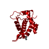 1ecwS C: citing same article ( S: Starting model for refinement |
|---|---|
| Similar structure data |


-Links
 PDBj
PDBj
- Assembly
| Deposited unit | 
| ||||||||
|---|---|---|---|---|---|---|---|---|---|
| 1 |
| ||||||||
| Unit cell |
|
-Components
| #1: Protein | Mass: 16358.727 Da / Num. of mol.: 1 Source method: isolated from a genetically manipulated source Source: (gene. exp.) Feline immunodeficiency virus / Strain: Petaluma / Gene: FIV Gag, gag / Plasmid: pRSET-B / Production host:  | ||
|---|---|---|---|
| #2: Chemical | ChemComp-EDO /   Mass: 62.068 Da / Num. of mol.: 4 / Source method: obtained synthetically / Formula: C2H6O2 Mass: 62.068 Da / Num. of mol.: 4 / Source method: obtained synthetically / Formula: C2H6O2#3: Water | ChemComp-HOH / |  Mass: 18.015 Da / Num. of mol.: 28 / Source method: isolated from a natural source / Formula: H2O Mass: 18.015 Da / Num. of mol.: 28 / Source method: isolated from a natural source / Formula: H2O |
-Experimental details
-Experiment
| Experiment | Method: X-RAY DIFFRACTION / Number of used crystals: 1 |
|---|
- Sample preparation
| Crystal grow | Temperature: 292 K / Method: vapor diffusion, sitting drop / pH: 6 Details: 0.1 M sodium acetate, 25% (w/v) polyethylene glycol 3,000, pH 6.0, VAPOR DIFFUSION, SITTING DROP, temperature 292K |
|---|
-Data collection
| Diffraction | Mean temperature: 100 K |
|---|---|
| Diffraction source | Source: SYNCHROTRON / Site: SOLEIL  / Beamline: PROXIMA 1 / Wavelength: 0.98011 Å / Beamline: PROXIMA 1 / Wavelength: 0.98011 Å |
| Detector | Type: PSI PILATUS 6M / Detector: PIXEL / Date: Feb 23, 2012 |
| Radiation | Monochromator: Kirkpatrick-Baez pair of bi-morph mirrors + channel-cut cryo-cooled silicon monochromator Protocol: SINGLE WAVELENGTH / Monochromatic (M) / Laue (L): M / Scattering type: x-ray |
| Radiation wavelength | Wavelength: 0.98011 Å / Relative weight: 1 |
| Reflection | Resolution: 2→20 Å / Num. obs: 7647 / % possible obs: 99.8 % / Observed criterion σ(F): 0 / Observed criterion σ(I): -3 / Redundancy: 6.9 % |
| Reflection shell | Resolution: 2→2.05 Å / Mean I/σ(I) obs: 4.78 / Num. unique all: 544 / % possible all: 99.8 |
- Processing
| Software |
| ||||||||||||||||||||||||||||
|---|---|---|---|---|---|---|---|---|---|---|---|---|---|---|---|---|---|---|---|---|---|---|---|---|---|---|---|---|---|
| Refinement | Method to determine structure: MOLECULAR REPLACEMENT Starting model: PDB entry 1ECW, SIV matrix protein Resolution: 2→19.31 Å / SU ML: 0.21 / σ(F): 2.01 / Phase error: 24.85 / Stereochemistry target values: Engh & Huber
| ||||||||||||||||||||||||||||
| Solvent computation | Shrinkage radii: 0.9 Å / VDW probe radii: 1.11 Å / Solvent model: FLAT BULK SOLVENT MODEL | ||||||||||||||||||||||||||||
| Refinement step | Cycle: LAST / Resolution: 2→19.31 Å
| ||||||||||||||||||||||||||||
| Refine LS restraints |
| ||||||||||||||||||||||||||||
| LS refinement shell |
|
