 Movie
Movie Controller

[English] 日本語
 Yorodumi
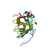
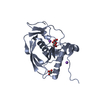
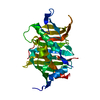
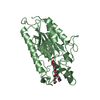
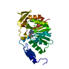
- PDB-1p6d: STRUCTURE OF THE D55N MUTANT OF PHOSPHOLIPASE C FROM BACILLUS CER... -
+ Open data

- Basic information
| Entry | Database: PDB / ID: 1p6d | ||||||
|---|---|---|---|---|---|---|---|
| Title | STRUCTURE OF THE D55N MUTANT OF PHOSPHOLIPASE C FROM BACILLUS CEREUS IN COMPLEX WITH (3S)-3,4,DI-N-HEXANOYLOXYBUTYL-1-PHOSPHOCHOLINE | ||||||
 Components Components | PHOSPHOLIPASE C | ||||||
 Keywords Keywords | HYDROLASE / TRI ZN2+ METAL CORE | ||||||
| Function / homology |  Function and homology information Function and homology informationphospholipase C / phosphatidylcholine phospholipase C activity / killing of cells of another organism / zinc ion binding Similarity search - Function | ||||||
| Biological species |  | ||||||
| Method |  X-RAY DIFFRACTION / FOURIER SYNTHESIS / Resolution: 2 Å X-RAY DIFFRACTION / FOURIER SYNTHESIS / Resolution: 2 Å | ||||||
 Authors Authors | Antikainen, N.M. / Monzingo, A.F. / Franklin, C.L. / Robertus, J.D. / Martin, S.F. | ||||||
 Citation Citation | Journal: Arch.Biochem.Biophys. / Year: 2003 Title: Using X-ray crystallography of the Asp55Asn mutant of the phosphatidylcholine-preferring phospholipase C from Bacillus cereus to support the mechanistic role of Asp55 as the general base. Authors: Antikainen, N.M. / Monzingo, A.F. / Franklin, C.L. / Robertus, J.D. / Martin, S.F. | ||||||
| History |
|
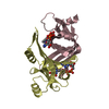
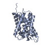
- Structure visualization
| Structure viewer | Molecule: MolmilJmol/JSmol |
|---|
- Downloads & links
-Download
| PDBx/mmCIF format | 1p6d.cif.gz | 65.6 KB | Display | PDBx/mmCIF format |
|---|---|---|---|---|
| PDB format | pdb1p6d.ent.gz | 47.7 KB | Display | PDB format |
| PDBx/mmJSON format | 1p6d.json.gz | Tree view | PDBx/mmJSON format | |
| Others |  Other downloads Other downloads |
-Validation report
| Summary document | 1p6d_validation.pdf.gz | 752.2 KB | Display | wwPDB validaton report |
|---|---|---|---|---|
| Full document | 1p6d_full_validation.pdf.gz | 758.3 KB | Display | |
| Data in XML | 1p6d_validation.xml.gz | 13.2 KB | Display | |
| Data in CIF | 1p6d_validation.cif.gz | 17.4 KB | Display | |
| Arichive directory | https://data.pdbj.org/pub/pdb/validation_reports/p6/1p6dftp://data.pdbj.org/pub/pdb/validation_reports/p6/1p6d | HTTPS FTP |
-Related structure data
| Related structure data |  1p5xC 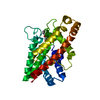 1p6eC 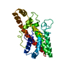 1ah7S S: Starting model for refinement C: citing same article ( |
|---|---|
| Similar structure data |


-Links
 PDBj
PDBj- Assembly
| Deposited unit | 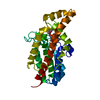
| ||||||||
|---|---|---|---|---|---|---|---|---|---|
| 1 |
| ||||||||
| Unit cell |
|
-Components
| #1: Protein | Mass: 28422.436 Da / Num. of mol.: 1 / Mutation: D55N Source method: isolated from a genetically manipulated source Source: (gene. exp.) | ||||
|---|---|---|---|---|---|
| #2: Chemical |   Mass: 65.409 Da / Num. of mol.: 3 / Source method: obtained synthetically / Formula: Zn Mass: 65.409 Da / Num. of mol.: 3 / Source method: obtained synthetically / Formula: Zn#3: Chemical | ChemComp-3PC / ( | 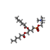  Mass: 451.534 Da / Num. of mol.: 1 / Source method: obtained synthetically / Formula: C21H42NO7P Mass: 451.534 Da / Num. of mol.: 1 / Source method: obtained synthetically / Formula: C21H42NO7P#4: Water | ChemComp-HOH / |  Mass: 18.015 Da / Num. of mol.: 86 / Source method: isolated from a natural source / Formula: H2O Mass: 18.015 Da / Num. of mol.: 86 / Source method: isolated from a natural source / Formula: H2O |
-Experimental details
-Experiment
| Experiment | Method: X-RAY DIFFRACTION / Number of used crystals: 1 |
|---|
- Sample preparation
| Crystal | Density Matthews: 2.63 Å3/Da / Density % sol: 53.22 % | ||||||||||||||||||||
|---|---|---|---|---|---|---|---|---|---|---|---|---|---|---|---|---|---|---|---|---|---|
| Crystal grow | Temperature: 298 K / Method: vapor diffusion, sitting drop / pH: 6 Details: AMMOUNIUM SULPHATE, pH 6, VAPOR DIFFUSION, SITTING DROP, temperature 298K | ||||||||||||||||||||
| Crystal grow | *PLUS Method: vapor diffusion | ||||||||||||||||||||
| Components of the solutions | *PLUS
|
-Data collection
| Diffraction | Mean temperature: 298 K |
|---|---|
| Diffraction source | Source: ROTATING ANODE / Type: RIGAKU RU200 / Wavelength: 1.5418 Å |
| Detector | Type: RIGAKU RAXIS IV / Detector: IMAGE PLATE / Date: May 18, 2000 / Details: MIRRORS |
| Radiation | Monochromator: DOUBLE FOCUSING MIRRORS (NI&PT) +NI FILTER / Protocol: SINGLE WAVELENGTH / Monochromatic (M) / Laue (L): M / Scattering type: x-ray |
| Radiation wavelength | Wavelength: 1.5418 Å / Relative weight: 1 |
| Reflection | Resolution: 2→20 Å / Num. all: 20598 / Num. obs: 20598 / % possible obs: 99.3 % / Observed criterion σ(F): 0 / Observed criterion σ(I): 0 / Redundancy: 5 % / Rmerge(I) obs: 0.074 / Net I/σ(I): 23.7 |
| Reflection shell | Resolution: 2→2.07 Å / Redundancy: 3.6 % / Rmerge(I) obs: 0.181 / % possible all: 98.3 |
| Reflection | *PLUS |
| Reflection shell | *PLUS Mean I/σ(I) obs: 7.5 |
- Processing
| Software |
| |||||||||||||||||||||||||
|---|---|---|---|---|---|---|---|---|---|---|---|---|---|---|---|---|---|---|---|---|---|---|---|---|---|---|
| Refinement | Method to determine structure: FOURIER SYNTHESIS Starting model: PDB ENTRY 1AH7 Resolution: 2→20 Å / σ(F): 0 / σ(I): 0 / Stereochemistry target values: Engh & Huber
| |||||||||||||||||||||||||
| Refinement step | Cycle: LAST / Resolution: 2→20 Å
| |||||||||||||||||||||||||
| Refine LS restraints |
| |||||||||||||||||||||||||
| Refinement | *PLUS Rfactor Rfree: 0.2303 | |||||||||||||||||||||||||
| Solvent computation | *PLUS | |||||||||||||||||||||||||
| Displacement parameters | *PLUS |
