| 登録情報 | データベース: PDB / ID: 1p5t
|
|---|
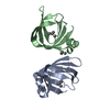
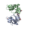
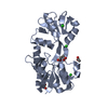
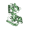
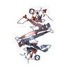
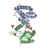
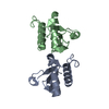
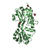
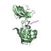
| タイトル | Crystal Structure of Dok1 PTB Domain |
|---|
 要素 要素 | Docking protein 1 |
|---|
 キーワード キーワード | SIGNALING PROTEIN |
|---|
| 機能・相同性 |  機能・相同性情報 機能・相同性情報
macrophage colony-stimulating factor signaling pathway / PTK6 Regulates RTKs and Their Effectors AKT1 and DOK1 / RET signaling / negative regulation of MAPK cascade / cell surface receptor protein tyrosine kinase signaling pathway / Ras protein signal transduction / intracellular signal transduction / nucleus / cytoplasm / cytosol類似検索 - 分子機能 Dok1/2/3, PTB domain / : / Phosphotyrosine-binding domain (IRS1-like) / IRS-type PTB domain profile. / Phosphotyrosine-binding domain / IRS-type PTB domain / PTB domain (IRS-1 type) / Pleckstrin-homology domain (PH domain)/Phosphotyrosine-binding domain (PTB) / PH-domain like / PH domain ...Dok1/2/3, PTB domain / : / Phosphotyrosine-binding domain (IRS1-like) / IRS-type PTB domain profile. / Phosphotyrosine-binding domain / IRS-type PTB domain / PTB domain (IRS-1 type) / Pleckstrin-homology domain (PH domain)/Phosphotyrosine-binding domain (PTB) / PH-domain like / PH domain / Pleckstrin homology domain. / Pleckstrin homology domain / PH-like domain superfamily / Roll / Mainly Beta類似検索 - ドメイン・相同性 |
|---|
| 生物種 |   Mus musculus (ハツカネズミ) Mus musculus (ハツカネズミ) |
|---|
| 手法 |  X線回折 / シンクロトロン / 多波長異常分散 / 解像度: 2.35 Å X線回折 / シンクロトロン / 多波長異常分散 / 解像度: 2.35 Å |
|---|
 データ登録者 データ登録者 | Shi, N. / Ye, S. / Liu, Y. / Zhou, W. / Ding, Y. / Lou, Z. / Qiang, B. / Yuan, J. / Rao, Z. |
|---|
 引用 引用 | ジャーナル: J.BIOL.CHEM. / 年: 2004
タイトル: Structural Basis for the Specific Recognition of RET by the Dok1 Phosphotyrosine Binding Domain
著者: Shi, N. / Ye, S. / Bartlam, M. / Yang, M. / Wu, J. / Liu, Y. / Sun, F. / Han, X. / Peng, X. / Qiang, B. / Yuan, J. / Rao, Z. |
|---|
| 履歴 | | 登録 | 2003年4月28日 | 登録サイト: RCSB / 処理サイト: PDBJ |
|---|
| 改定 1.0 | 2004年2月17日 | Provider: repository / タイプ: Initial release |
|---|
| 改定 1.1 | 2008年4月29日 | Group: Version format compliance |
|---|
| 改定 1.2 | 2011年7月13日 | Group: Version format compliance |
|---|
| 改定 1.3 | 2024年10月16日 | Group: Data collection / Database references ...Data collection / Database references / Derived calculations / Structure summary
カテゴリ: chem_comp_atom / chem_comp_bond ...chem_comp_atom / chem_comp_bond / database_2 / pdbx_entry_details / pdbx_modification_feature / struct_conn / struct_ref_seq_dif
Item: _database_2.pdbx_DOI / _database_2.pdbx_database_accession ..._database_2.pdbx_DOI / _database_2.pdbx_database_accession / _struct_conn.pdbx_leaving_atom_flag / _struct_ref_seq_dif.details |
|---|
|
|---|
 ムービー
ムービー コントローラー


 データを開く
 基本情報
基本情報 構造の表示
構造の表示 ダウンロードとリンク
 その他のダウンロード

 PDBj
PDBj


 集合体
集合体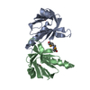

 分子量: 18.015 Da / 分子数: 16 / 由来タイプ: 天然 / 式: H2O
 試料調製
試料調製 / ビームライン: BL41XU / 波長: 0.9798, 0.9800, 0.9000
 解析
解析