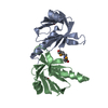
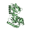
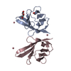
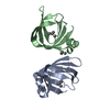
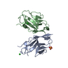
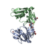
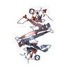
[English] 日本語
Yorodumi
- PDB-4u17: Swapped dimer of the human Fyn-SH2 domain -

+
Open data


ID or keywords:

Loading...

-
Basic information

Entry
Database: PDB / ID: 4u17
TitleSwapped dimer of the human Fyn-SH2 domain
ComponentsTyrosine-protein kinase Fyn
KeywordsTRANSFERASE / human Fyn-SH2 domain / domain swapping
Function / homology
Function and homology information


response to singlet oxygen / Reelin signalling pathway / negative regulation of hydrogen peroxide biosynthetic process / perinuclear endoplasmic reticulum / NTRK2 activates RAC1 / growth factor receptor binding / Activated NTRK2 signals through FYN / heart process / cellular response to L-glutamate / SEMA3A-Plexin repulsion signaling by inhibiting Integrin adhesion ...response to singlet oxygen / Reelin signalling pathway / negative regulation of hydrogen peroxide biosynthetic process / perinuclear endoplasmic reticulum / NTRK2 activates RAC1 / growth factor receptor binding / Activated NTRK2 signals through FYN / heart process / cellular response to L-glutamate / SEMA3A-Plexin repulsion signaling by inhibiting Integrin adhesion / regulation of glutamate receptor signaling pathway / regulation of calcium ion import across plasma membrane / Platelet Adhesion to exposed collagen / G protein-coupled glutamate receptor signaling pathway / CD28 co-stimulation / positive regulation of protein localization to membrane / activated T cell proliferation / CRMPs in Sema3A signaling / positive regulation of cysteine-type endopeptidase activity / FLT3 signaling through SRC family kinases / negative regulation of dendritic spine maintenance / feeding behavior / Nef and signal transduction / type 5 metabotropic glutamate receptor binding / Nephrin family interactions / dendrite morphogenesis / DCC mediated attractive signaling / EPH-Ephrin signaling / CD28 dependent Vav1 pathway / Ephrin signaling / dendritic spine maintenance / Regulation of KIT signaling / tau-protein kinase activity / CTLA4 inhibitory signaling / phospholipase activator activity / leukocyte migration / Fc-gamma receptor signaling pathway involved in phagocytosis / EPHA-mediated growth cone collapse / cellular response to platelet-derived growth factor stimulus / Dectin-2 family / stimulatory C-type lectin receptor signaling pathway / PECAM1 interactions / glial cell projection / CD28 dependent PI3K/Akt signaling / phospholipase binding / response to amyloid-beta / Sema3A PAK dependent Axon repulsion / cellular response to glycine / FCGR activation / alpha-tubulin binding / EPH-ephrin mediated repulsion of cells / positive regulation of protein targeting to membrane / ephrin receptor signaling pathway / Role of LAT2/NTAL/LAB on calcium mobilization / vascular endothelial growth factor receptor signaling pathway / detection of mechanical stimulus involved in sensory perception of pain / negative regulation of oxidative stress-induced intrinsic apoptotic signaling pathway / regulation of peptidyl-tyrosine phosphorylation / positive regulation of tyrosine phosphorylation of STAT protein / forebrain development / negative regulation of inflammatory response to antigenic stimulus / extrinsic component of cytoplasmic side of plasma membrane / Signaling by ERBB2 / negative regulation of protein ubiquitination / GPVI-mediated activation cascade / cellular response to transforming growth factor beta stimulus / T cell costimulation / EPHB-mediated forward signaling / ephrin receptor binding / NCAM signaling for neurite out-growth / CD209 (DC-SIGN) signaling / FCGR3A-mediated IL10 synthesis / Signaling by phosphorylated juxtamembrane, extracellular and kinase domain KIT mutants / Antigen activates B Cell Receptor (BCR) leading to generation of second messengers / learning / cell surface receptor protein tyrosine kinase signaling pathway / Regulation of signaling by CBL / actin filament / Cell surface interactions at the vascular wall / FCGR3A-mediated phagocytosis / axon guidance / non-specific protein-tyrosine kinase / neuron migration / non-membrane spanning protein tyrosine kinase activity / Schaffer collateral - CA1 synapse / protein catabolic process / modulation of chemical synaptic transmission / tau protein binding / Signaling by SCF-KIT / negative regulation of protein catabolic process / positive regulation of neuron projection development / VEGFA-VEGFR2 Pathway / cellular response to hydrogen peroxide / positive regulation of protein localization to nucleus / peptidyl-tyrosine phosphorylation / cellular response to amyloid-beta / calcium ion transport / Constitutive Signaling by Aberrant PI3K in Cancer / Signaling by CSF1 (M-CSF) in myeloid cells / disordered domain specific binding
Similarity search - Function
: / Fyn/Yrk, SH3 domain / SH2 domain / SHC Adaptor Protein / SH3 domain / SH2 domain / Src homology 2 (SH2) domain profile. / Src homology 2 domains / SH2 domain / Src homology 3 domains ...: / Fyn/Yrk, SH3 domain / SH2 domain / SHC Adaptor Protein / SH3 domain / SH2 domain / Src homology 2 (SH2) domain profile. / Src homology 2 domains / SH2 domain / Src homology 3 domains / SH2 domain superfamily / SH3-like domain superfamily / Src homology 3 (SH3) domain profile. / SH3 domain / Tyrosine-protein kinase, catalytic domain / Tyrosine kinase, catalytic domain / Tyrosine protein kinases specific active-site signature. / Tyrosine-protein kinase, active site / Protein tyrosine and serine/threonine kinase / Serine-threonine/tyrosine-protein kinase, catalytic domain / Protein kinase, ATP binding site / Protein kinases ATP-binding region signature. / Protein kinase domain profile. / Protein kinase domain / Protein kinase-like domain superfamily / 2-Layer Sandwich / Alpha Beta
Similarity search - Domain/homology
PHOSPHATE ION / Tyrosine-protein kinase Fyn
Similarity search - Component
Biological speciesHomo sapiens (human)
MethodX-RAY DIFFRACTION / SYNCHROTRON / MOLECULAR REPLACEMENT / Resolution: 1.99 Å
AuthorsGarcia-Pino, A. / Huculeci, R. / Lenaerts, T. / van Nuland, N.A.J.
CitationJournal: To Be Published
Title: Swapped dimer of the human Fyn-SH2 domain
Authors: Garcia-Pino, A. / Huculeci, R. / Lenaerts, T. / van Nuland, N.A.J.
History
DepositionJul 15, 2014Deposition site: RCSB / Processing site: PDBE
Revision 1.0Jul 29, 2015Provider: repository / Type: Initial release
Revision 1.1Dec 20, 2023Group: Data collection / Database references / Refinement description
Category: chem_comp_atom / chem_comp_bond ...chem_comp_atom / chem_comp_bond / database_2 / pdbx_initial_refinement_model
Item: _database_2.pdbx_DOI / _database_2.pdbx_database_accession

-
Structure visualization

Structure viewerMolecule:
MolmilJmol/JSmol

Downloads & links

-
Assembly

Deposited unit
A: Tyrosine-protein kinase Fyn
B: Tyrosine-protein kinase Fyn
C: Tyrosine-protein kinase Fyn
hetero molecules


Theoretical massNumber of molelcules
Total (without water)37,61214
Polymers36,6993
Non-polymers91311
Water2,720151
1
A: Tyrosine-protein kinase Fyn
C: Tyrosine-protein kinase Fyn
hetero molecules


Theoretical massNumber of molelcules
Total (without water)25,12710
Polymers24,4662
Non-polymers6618
Water362
TypeNameSymmetry operationNumber
identity operation1_555x,y,z1
Buried area6620 Å2
ΔGint-71 kcal/mol
Surface area11250 Å2
MethodPISA
2
B: Tyrosine-protein kinase Fyn
hetero molecules

B: Tyrosine-protein kinase Fyn
hetero molecules


Theoretical massNumber of molelcules
Total (without water)24,9708
Polymers24,4662
Non-polymers5046
Water362
TypeNameSymmetry operationNumber
identity operation1_555x,y,z1
crystal symmetry operation2_555-x,y,-z1
Buried area6260 Å2
ΔGint-70 kcal/mol
Surface area10750 Å2
MethodPISA
Unit cell
Length a, b, c (Å)88.965, 57.873, 101.188
Angle α, β, γ (deg.)90.00, 90.60, 90.00
Int Tables number5
Space group name H-MC121

-
Components

#1: Protein Tyrosine-protein kinase Fyn / Proto-oncogene Syn / Proto-oncogene c-Fyn / Src-like kinase / SLK / p59-Fyn


Mass: 12232.927 Da / Num. of mol.: 3 / Fragment: Fyn-SH2 domain (UNP residues 148-248)
Source method: isolated from a genetically manipulated source
Source: (gene. exp.) Homo sapiens (human) / Gene: FYN / Production host: Escherichia coli BL21(DE3) (bacteria)
References: UniProt: P06241, non-specific protein-tyrosine kinase
#2: Chemical
ChemComp-PO4 / PHOSPHATE ION


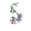
Mass: 94.971 Da / Num. of mol.: 7 / Source method: obtained synthetically / Formula: PO4
#3: Chemical
ChemComp-EDO / 1,2-ETHANEDIOL / ETHYLENE GLYCOL


Mass: 62.068 Da / Num. of mol.: 4 / Source method: obtained synthetically / Formula: C2H6O2
#4: Water ChemComp-HOH / water


Mass: 18.015 Da / Num. of mol.: 151 / Source method: isolated from a natural source / Formula: H2O

-
Experimental details

-
Experiment

ExperimentMethod: X-RAY DIFFRACTION

-
Sample preparation

CrystalDensity Matthews: 3.55 Å3/Da / Density % sol: 65.34 %
Crystal growTemperature: 293 K / Method: vapor diffusion, hanging drop / pH: 6.5 / Details: 50 mM sodium phosphate pH 6.5

-
Data collection

DiffractionMean temperature: 100 K
Diffraction sourceSource: SYNCHROTRON / Site: SOLEIL / Beamline: PROXIMA 1 / Wavelength: 0.9801 Å
DetectorType: ADSC QUANTUM 315r / Detector: CCD / Date: Jul 4, 2011
RadiationProtocol: SINGLE WAVELENGTH / Monochromatic (M) / Laue (L): M / Scattering type: x-ray
Radiation wavelengthWavelength: 0.9801 Å / Relative weight: 1
ReflectionResolution: 1.99→34.92 Å / Num. obs: 35049 / % possible obs: 98.66 % / Redundancy: 3.6 % / Rsym value: 0.081 / Net I/σ(I): 7.7
Reflection shellResolution: 1.99→2.1 Å / Redundancy: 3.6 % / Rmerge(I) obs: 0.303 / Mean I/σ(I) obs: 3.2 / % possible all: 98.9

-
Processing

Software
NameVersionClassification
PHENIX(phenix.refine: 1.9_1692)refinement
iMOSFLMdata reduction
SCALAdata scaling
RefinementMethod to determine structure: MOLECULAR REPLACEMENT
Starting model: 1G83
Resolution: 1.99→34.916 Å / SU ML: 0.29 / Cross valid method: THROUGHOUT / σ(F): 1.36 / Phase error: 31.91 / Stereochemistry target values: ML
RfactorNum. reflection% reflectionSelection details
Rfree0.2554 1743 4.98 %Random selection
Rwork0.2068 ---
obs0.2093 34985 98.48 %-
Solvent computationShrinkage radii: 0.9 Å / VDW probe radii: 1.11 Å / Solvent model: FLAT BULK SOLVENT MODEL
Refinement stepCycle: LAST / Resolution: 1.99→34.916 Å
ProteinNucleic acidLigandSolventTotal
Num. atoms2434 0 51 151 2636
Refine LS restraints
Refine-IDTypeDev idealNumber
X-RAY DIFFRACTIONf_bond_d0.0122539
X-RAY DIFFRACTIONf_angle_d1.4133424
X-RAY DIFFRACTIONf_dihedral_angle_d15.607900
X-RAY DIFFRACTIONf_chiral_restr0.057353
X-RAY DIFFRACTIONf_plane_restr0.006433
LS refinement shell
Resolution (Å)Rfactor RfreeNum. reflection RfreeRfactor RworkNum. reflection RworkRefine-ID% reflection obs (%)
1.9895-2.04810.32981440.312624X-RAY DIFFRACTION94
2.0481-2.11420.35451470.29592779X-RAY DIFFRACTION99
2.1142-2.18970.30341260.27082759X-RAY DIFFRACTION99
2.1897-2.27740.311400.25492773X-RAY DIFFRACTION99
2.2774-2.3810.29851370.25542789X-RAY DIFFRACTION99
2.381-2.50650.2661270.24232801X-RAY DIFFRACTION99
2.5065-2.66350.31681500.24212794X-RAY DIFFRACTION99
2.6635-2.8690.31091710.2332740X-RAY DIFFRACTION99
2.869-3.15760.29971560.23022764X-RAY DIFFRACTION99
3.1576-3.61410.25711570.18592782X-RAY DIFFRACTION99
3.6141-4.55180.19451470.16662769X-RAY DIFFRACTION98
4.5518-34.92120.22151410.1832868X-RAY DIFFRACTION98
Refinement TLS params.

Method: refined / Refine-ID: X-RAY DIFFRACTION

IDL112)L122)L132)L222)L232)L332)S11 (Å °)S12 (Å °)S13 (Å °)S21 (Å °)S22 (Å °)S23 (Å °)S31 (Å °)S32 (Å °)S33 (Å °)T112)T122)T132)T222)T232)T332)Origin x (Å)Origin y (Å)Origin z (Å)
13.8941.28972.95168.166-3.03024.86470.3612-1.1157-0.97730.6195-0.75620.18040.78331.2021-0.15650.69480.06190.06790.55410.1450.457517.3157-10.886943.8886
22.0522-0.62120.70176.10470.96858.11150.03930.1935-0.25340.1465-0.0626-0.28660.36390.6839-0.0060.2743-0.0793-0.02210.4955-0.02060.321517.4158-6.421332.9794
32.485-0.80681.16296.4456-2.68737.06940.227-0.52810.0898-0.1931-0.05990.01370.0039-0.55-0.20750.4106-0.1975-0.00960.5555-0.03690.26124.567814.372430.3981
43.5487-1.0726-3.39773.3312.06069.20050.1949-0.40380.3531-0.1373-0.0122-0.2719-0.19010.6205-0.21160.5688-0.0760.03830.33960.00220.284818.1148-3.30921.6035
55.0522-4.6725-4.89339.4227.7258.13060.45060.55030.4933-0.6236-0.2511-0.6718-0.2881-0.6577-0.27180.59950.0523-0.02280.30910.01320.313211.7031-0.7616-0.3356
65.98732.53576.3073.22192.32017.79290.19890.2851-0.10070.01270.2066-0.0490.32750.1585-0.49390.42530.00450.02680.60040.01620.26081.00831.29410.6794
77.89953.72940.55756.0309-2.11826.4172-0.51610.3648-0.7623-0.25160.2786-0.30361.24790.64450.30850.68540.13160.05230.4216-0.0170.3165-11.2054-9.6449-7.0086
84.5738-1.41021.28125.70052.5965.2139-0.098-0.23480.4977-0.2230.1772-0.4835-1.26420.2368-0.0020.5321-0.1041-0.01990.3203-0.06090.34430.22924.601232.2444
90.5022-0.6993-0.71512.7401-1.07913.42380.4991-0.43430.00220.5903-0.34590.0475-0.87820.2369-0.04760.6179-0.2316-0.0580.5076-0.00450.313420.12793.737637.4098
Refinement TLS group
IDRefine-IDRefine TLS-IDSelection details
1X-RAY DIFFRACTION1chain 'A' and (resseq 11:19)
2X-RAY DIFFRACTION2chain 'A' and (resseq 20:71)
3X-RAY DIFFRACTION3chain 'A' and (resseq 72:112)
4X-RAY DIFFRACTION4chain 'B' and (resseq 11:60)
5X-RAY DIFFRACTION5chain 'B' and (resseq 61:71)
6X-RAY DIFFRACTION6chain 'B' and (resseq 72:87)
7X-RAY DIFFRACTION7chain 'B' and (resseq 88:112)
8X-RAY DIFFRACTION8chain 'C' and (resseq 10:71)
9X-RAY DIFFRACTION9chain 'C' and (resseq 72:112)

+
About Yorodumi

-
News

-
Feb 9, 2022. New format data for meta-information of EMDB entries

New format data for meta-information of EMDB entries

  • Version 3 of the EMDB header file is now the official format.
  • The previous official version 1.9 will be removed from the archive.

Related info.:EMDB header

External links:wwPDB to switch to version 3 of the EMDB data model

-
Aug 12, 2020. Covid-19 info

Covid-19 info

URL: https://pdbjlvh1.pdbj.org/emnavi/covid19.php

New page: Covid-19 featured information page in EM Navigator.

Related info.:Covid-19 info / Mar 5, 2020. Novel coronavirus structure data

+
Mar 5, 2020. Novel coronavirus structure data

Novel coronavirus structure data

Related info.:Yorodumi Speices / Aug 12, 2020. Covid-19 info

External links:COVID-19 featured content - PDBj / Molecule of the Month (242):Coronavirus Proteases

+
Jan 31, 2019. EMDB accession codes are about to change! (news from PDBe EMDB page)

EMDB accession codes are about to change! (news from PDBe EMDB page)

  • The allocation of 4 digits for EMDB accession codes will soon come to an end. Whilst these codes will remain in use, new EMDB accession codes will include an additional digit and will expand incrementally as the available range of codes is exhausted. The current 4-digit format prefixed with “EMD-” (i.e. EMD-XXXX) will advance to a 5-digit format (i.e. EMD-XXXXX), and so on. It is currently estimated that the 4-digit codes will be depleted around Spring 2019, at which point the 5-digit format will come into force.
  • The EM Navigator/Yorodumi systems omit the EMD- prefix.

Related info.:Q: What is EMD? / ID/Accession-code notation in Yorodumi/EM Navigator

External links:EMDB Accession Codes are Changing Soon! / Contact to PDBj

+
Jul 12, 2017. Major update of PDB

Major update of PDB

  • wwPDB released updated PDB data conforming to the new PDBx/mmCIF dictionary.
  • This is a major update changing the version number from 4 to 5, and with Remediation, in which all the entries are updated.
  • In this update, many items about electron microscopy experimental information are reorganized (e.g. em_software).
  • Now, EM Navigator and Yorodumi are based on the updated data.

External links:wwPDB Remediation / Enriched Model Files Conforming to OneDep Data Standards Now Available in the PDB FTP Archive

-
Yorodumi

Thousand views of thousand structures

  • Yorodumi is a browser for structure data from EMDB, PDB, SASBDB, etc.
  • This page is also the successor to EM Navigator detail page, and also detail information page/front-end page for Omokage search.
  • The word "yorodu" (or yorozu) is an old Japanese word meaning "ten thousand". "mi" (miru) is to see.

Related info.:EMDB / PDB / SASBDB / Comparison of 3 databanks / Yorodumi Search / Aug 31, 2016. New EM Navigator & Yorodumi / Yorodumi Papers / Jmol/JSmol / Function and homology information / Changes in new EM Navigator and Yorodumi

Read more