 Movie
Movie Controller

[English] 日本語
 Yorodumi
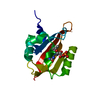
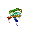
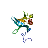
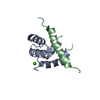
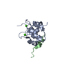
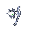
- PDB-1oqp: STRUCTURE OF THE CA2+/C-TERMINAL DOMAIN OF CALTRACTIN IN COMPLEX ... -
+ Open data

- Basic information
| Entry | Database: PDB / ID: 1oqp | ||||||
|---|---|---|---|---|---|---|---|
| Title | STRUCTURE OF THE CA2+/C-TERMINAL DOMAIN OF CALTRACTIN IN COMPLEX WITH THE CDC31P-BINDING DOMAIN FROM KAR1P | ||||||
 Components Components |
| ||||||
 Keywords Keywords | PROTEIN BINDING / protein-peptide complex / caltractin / KAR1P / calcium-binding | ||||||
| Function / homology |  Function and homology information Function and homology informationhalf bridge of spindle pole body / spindle pole body duplication / karyogamy involved in conjugation with cellular fusion / inner dynein arm / dynein heavy chain binding / microtubule / cell division / calcium ion binding / endoplasmic reticulum Similarity search - Function | ||||||
| Biological species |   Chlamydomonas reinhardtii (plant) Chlamydomonas reinhardtii (plant) | ||||||
| Method | SOLUTION NMR / torsion angle dynamics | ||||||
 Authors Authors | Hu, H.T. / Chazin, W.J. | ||||||
 Citation Citation | Journal: J.Mol.Biol. / Year: 2003 Title: Unique Features in the C-terminal Domain Provide Caltractin with Target Specificity Authors: Hu, H.T. / Chazin, W.J. | ||||||
| History |
|
- Structure visualization
| Structure viewer | Molecule: MolmilJmol/JSmol |
|---|
- Downloads & links
-Download
| PDBx/mmCIF format | 1oqp.cif.gz | 616.5 KB | Display | PDBx/mmCIF format |
|---|---|---|---|---|
| PDB format | pdb1oqp.ent.gz | 516.4 KB | Display | PDB format |
| PDBx/mmJSON format | 1oqp.json.gz | Tree view | PDBx/mmJSON format | |
| Others |  Other downloads Other downloads |
-Validation report
| Arichive directory | https://data.pdbj.org/pub/pdb/validation_reports/oq/1oqpftp://data.pdbj.org/pub/pdb/validation_reports/oq/1oqp | HTTPS FTP |
|---|
-Related structure data
| Similar structure data |
|---|


-Links
 PDBj
PDBj- Assembly
| Deposited unit | 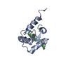
| |||||||||
|---|---|---|---|---|---|---|---|---|---|---|
| 1 |
| |||||||||
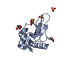
| NMR ensembles |
|
-Components
| #1: Protein | Mass: 8977.838 Da / Num. of mol.: 1 Source method: isolated from a genetically manipulated source Source: (gene. exp.) Chlamydomonas reinhardtii (plant) / Plasmid: PET / Production host:  |
|---|---|
| #2: Protein/peptide | Mass: 2501.003 Da / Num. of mol.: 1 / Source method: obtained synthetically Details: The peptide consists of the cdc31p-binding domain from Kar1p, residues 239-257, and has been chemically synthesized using solid phase F-Moc chemistry. The sequence is naturally found in ...Details: The peptide consists of the cdc31p-binding domain from Kar1p, residues 239-257, and has been chemically synthesized using solid phase F-Moc chemistry. The sequence is naturally found in Saccharomyces cerevisiae (baker's yeast). References: UniProt: P11927 |
-Experimental details
-Experiment
| Experiment | Method: SOLUTION NMR | ||||||||||||||||||||||||
|---|---|---|---|---|---|---|---|---|---|---|---|---|---|---|---|---|---|---|---|---|---|---|---|---|---|
| NMR experiment |
|
- Sample preparation
| Details |
| ||||||||||||
|---|---|---|---|---|---|---|---|---|---|---|---|---|---|
| Sample conditions | Ionic strength: 50mM KCl, 5mM CaCl2 / pH: 7 / Pressure: ambient / Temperature: 298 K | ||||||||||||
| Crystal grow | *PLUS Method: other / Details: NMR |
-NMR measurement
| Radiation | Protocol: SINGLE WAVELENGTH / Monochromatic (M) / Laue (L): M | |||||||||||||||
|---|---|---|---|---|---|---|---|---|---|---|---|---|---|---|---|---|
| Radiation wavelength | Relative weight: 1 | |||||||||||||||
| NMR spectrometer |
|
- Processing
| NMR software |
| ||||||||||||||||||||||||
|---|---|---|---|---|---|---|---|---|---|---|---|---|---|---|---|---|---|---|---|---|---|---|---|---|---|
| Refinement | Method: torsion angle dynamics / Software ordinal: 1 Details: the structures are based on a total of 1827 constraints, 1641 are NOE-derived distance constraints, 134 dihedral angle constraints, 52 distance constraints from hydrogen bonds. | ||||||||||||||||||||||||
| NMR ensemble | Conformer selection criteria: structures with the least restraint violations,structures with the lowest energy Conformers calculated total number: 60 / Conformers submitted total number: 20 |
 Amber
Amber