 Movie
Movie Controller


+ Open data

- Basic information
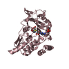
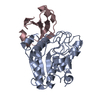
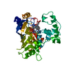
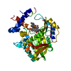
| Entry | Database: PDB / ID: 1ooc | ||||||
|---|---|---|---|---|---|---|---|
| Title | Mutations in the T1.5 loop of pectate lyase A | ||||||
 Components Components | Pectate lyase A | ||||||
 Keywords Keywords | LYASE / parallel beta helix | ||||||
| Function / homology |  Function and homology information Function and homology informationpectate lyase / pectate lyase activity / pectin catabolic process / extracellular region / metal ion binding Similarity search - Function | ||||||
| Biological species |  Erwinia chrysanthemi (bacteria) Erwinia chrysanthemi (bacteria) | ||||||
| Method |  X-RAY DIFFRACTION / MOLECULAR REPLACEMENT / Resolution: 2.94 Å X-RAY DIFFRACTION / MOLECULAR REPLACEMENT / Resolution: 2.94 Å | ||||||
 Authors Authors | Dehdashti, S.J. / Doan, C.N. / Chao, K. / Vordtriede, P.B. / Yoder, M.D. | ||||||
 Citation Citation | Journal: Acta Crystallogr.,Sect.D / Year: 2003 Title: Effect of mutations in the T1.5 loop of pectate lyase A from Erwinia chrysanthemi EC16. Authors: Dehdashti, S.J. / Doan, C.N. / Chao, K.L. / Yoder, M.D. | ||||||
| History |
|
- Structure visualization
| Structure viewer | Molecule: MolmilJmol/JSmol |
|---|
- Downloads & links
-Download
| PDBx/mmCIF format | 1ooc.cif.gz | 141.5 KB | Display | PDBx/mmCIF format |
|---|---|---|---|---|
| PDB format | pdb1ooc.ent.gz | 112.7 KB | Display | PDB format |
| PDBx/mmJSON format | 1ooc.json.gz | Tree view | PDBx/mmJSON format | |
| Others |  Other downloads Other downloads |
-Validation report
| Arichive directory | https://data.pdbj.org/pub/pdb/validation_reports/oo/1oocftp://data.pdbj.org/pub/pdb/validation_reports/oo/1ooc | HTTPS FTP |
|---|
-Related structure data
| Related structure data |  1pe9C  1jtaS S: Starting model for refinement C: citing same article ( |
|---|---|
| Similar structure data |


-Links
 PDBj
PDBj- Assembly
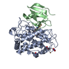
| Deposited unit | 
| ||||||||
|---|---|---|---|---|---|---|---|---|---|
| 1 | 
| ||||||||
| 2 | 
| ||||||||
| Unit cell |
| ||||||||
| Details | The biological assembly is a monomer that packs two molecules in the asymmetric unit. |
-Components
| #1: Protein | Mass: 38739.535 Da / Num. of mol.: 2 / Fragment: T1.5 / Mutation: N215S, T217S, S219G, A220S Source method: isolated from a genetically manipulated source Source: (gene. exp.) Erwinia chrysanthemi (bacteria) / Genus: Dickeya / Gene: PELA / Plasmid: pMDY29 / Production host: References: UniProt: P29155, UniProt: P0C1A2*PLUS, pectate lyase Has protein modification | Y | |
|---|
-Experimental details
-Experiment
| Experiment | Method: X-RAY DIFFRACTION / Number of used crystals: 1 |
|---|
- Sample preparation
| Crystal | Density Matthews: 2.39 Å3/Da / Density % sol: 48.04 % | ||||||||||||||||||||||||||||||||||||
|---|---|---|---|---|---|---|---|---|---|---|---|---|---|---|---|---|---|---|---|---|---|---|---|---|---|---|---|---|---|---|---|---|---|---|---|---|---|
| Crystal grow | Temperature: 293 K / Method: vapor diffusion, hanging drop / pH: 6.5 Details: PEG 5000 monomethyl ether (MME), 0.1M MES (2-[N-Morpholino]ethanesulfonic acid), pH 6.5, VAPOR DIFFUSION, HANGING DROP, temperature 293K | ||||||||||||||||||||||||||||||||||||
| Crystal grow | *PLUS Temperature: 295 K / Method: vapor diffusion, hanging drop | ||||||||||||||||||||||||||||||||||||
| Components of the solutions | *PLUS
|
-Data collection
| Diffraction | Mean temperature: 153 K |
|---|---|
| Diffraction source | Source: ROTATING ANODE / Type: RIGAKU RU200 / Wavelength: 1.5418 |
| Detector | Type: MAR scanner 300 mm plate / Detector: IMAGE PLATE / Date: Jan 24, 2003 / Details: Osmic mirrors |
| Radiation | Monochromator: Osmic mirrors / Protocol: SINGLE WAVELENGTH / Monochromatic (M) / Laue (L): M / Scattering type: x-ray |
| Radiation wavelength | Wavelength: 1.5418 Å / Relative weight: 1 |
| Reflection | Resolution: 2.9→50 Å / Num. all: 14754 / Num. obs: 14754 / % possible obs: 91.6 % / Observed criterion σ(F): 0 / Observed criterion σ(I): 0 / Redundancy: 8.7 % / Biso Wilson estimate: 47.22 Å2 / Rsym value: 0.097 / Net I/σ(I): 23.6 |
| Reflection shell | Resolution: 2.9→3.08 Å / Mean I/σ(I) obs: 14.6 / Num. unique all: 1511 / Rsym value: 0.273 / % possible all: 81 |
| Reflection | *PLUS Num. measured all: 128721 / Rmerge(I) obs: 0.097 |
| Reflection shell | *PLUS % possible obs: 81 % / Rmerge(I) obs: 0.273 / Mean I/σ(I) obs: 8.35 |
- Processing
| Software |
| |||||||||||||||||||||||||
|---|---|---|---|---|---|---|---|---|---|---|---|---|---|---|---|---|---|---|---|---|---|---|---|---|---|---|
| Refinement | Method to determine structure: MOLECULAR REPLACEMENT Starting model: PDB entry 1JTA with out residues 215-226 Resolution: 2.94→29.55 Å / Rfactor Rfree error: 0.01 / Isotropic thermal model: RESTRAINED / Cross valid method: THROUGHOUT / σ(F): 0 / σ(I): 0 / Stereochemistry target values: Engh & Huber
| |||||||||||||||||||||||||
| Solvent computation | Solvent model: FLAT MODEL / Bsol: 10 Å2 / ksol: 0.372263 e/Å3 | |||||||||||||||||||||||||
| Displacement parameters | Biso mean: 17.4 Å2
| |||||||||||||||||||||||||
| Refine analyze |
| |||||||||||||||||||||||||
| Refinement step | Cycle: LAST / Resolution: 2.94→29.55 Å
| |||||||||||||||||||||||||
| Refine LS restraints |
| |||||||||||||||||||||||||
| Refine LS restraints NCS | NCS model details: CONSTR | |||||||||||||||||||||||||
| LS refinement shell | Resolution: 2.94→3.08 Å / Rfactor Rfree error: 0.034 / Total num. of bins used: 6
| |||||||||||||||||||||||||
| Xplor file | Serial no: 1 / Param file: PROTEIN_REP.PARAM / Topol file: PROTEIN.TOP | |||||||||||||||||||||||||
| Refinement | *PLUS Highest resolution: 2.9 Å / Lowest resolution: 50 Å / Num. reflection Rfree: 1511 / Rfactor Rfree: 0.2748 / Rfactor Rwork: 0.2195 | |||||||||||||||||||||||||
| Solvent computation | *PLUS | |||||||||||||||||||||||||
| Displacement parameters | *PLUS | |||||||||||||||||||||||||
| Refine LS restraints | *PLUS
|
