 Movie
Movie Controller

[English] 日本語
 Yorodumi
- PDB-1n6j: Structural basis of sequence-specific recruitment of histone deac... -
+ Open data

- Basic information
| Entry | Database: PDB / ID: 1n6j | ||||||
|---|---|---|---|---|---|---|---|
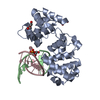
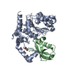
| Title | Structural basis of sequence-specific recruitment of histone deacetylases by Myocyte Enhancer Factor-2 | ||||||
 Components Components |
| ||||||
 Keywords Keywords | TRANSCRIPTION/DNA / MADS-box / Protein-DNA complex / histone deacetylases / TRANSCRIPTION-DNA COMPLEX | ||||||
| Function / homology |  Function and homology information Function and homology informationregulation of chromatin organization / muscle organ development / Formation of Senescence-Associated Heterochromatin Foci (SAHF) / Myogenesis / protein phosphatase inhibitor activity / histone deacetylase binding / cell junction / sequence-specific double-stranded DNA binding / transcription corepressor activity / heart development ...regulation of chromatin organization / muscle organ development / Formation of Senescence-Associated Heterochromatin Foci (SAHF) / Myogenesis / protein phosphatase inhibitor activity / histone deacetylase binding / cell junction / sequence-specific double-stranded DNA binding / transcription corepressor activity / heart development / nucleosome assembly / chromatin organization / DNA-binding transcription activator activity, RNA polymerase II-specific / transcription regulator complex / DNA-binding transcription factor activity, RNA polymerase II-specific / cell differentiation / cell surface receptor signaling pathway / protein dimerization activity / RNA polymerase II cis-regulatory region sequence-specific DNA binding / DNA-binding transcription factor activity / chromatin / negative regulation of transcription by RNA polymerase II / positive regulation of transcription by RNA polymerase II / nucleoplasm / nucleus / cytosol Similarity search - Function | ||||||
| Biological species |  Homo sapiens (human) Homo sapiens (human) | ||||||
| Method |  X-RAY DIFFRACTION / SYNCHROTRON / MOLECULAR REPLACEMENT / Resolution: 2.2 Å X-RAY DIFFRACTION / SYNCHROTRON / MOLECULAR REPLACEMENT / Resolution: 2.2 Å | ||||||
 Authors Authors | Han, A. / Pan, F. / Stroud, J.C. / Youn, H.D. / Liu, J.O. / Chen, L. | ||||||
 Citation Citation | Journal: Nature / Year: 2003 Title: Sequence-specific recruitment of transcriptional co-repressor Cabin1 by myocyte enhancer factor-2 Authors: Han, A. / Pan, F. / Stroud, J.C. / Youn, H.D. / Liu, J.O. / Chen, L. | ||||||
| History |
|
- Structure visualization
| Structure viewer | Molecule: MolmilJmol/JSmol |
|---|
- Downloads & links
-Download
| PDBx/mmCIF format | 1n6j.cif.gz | 131.7 KB | Display | PDBx/mmCIF format |
|---|---|---|---|---|
| PDB format | pdb1n6j.ent.gz | 103.3 KB | Display | PDB format |
| PDBx/mmJSON format | 1n6j.json.gz | Tree view | PDBx/mmJSON format | |
| Others |  Other downloads Other downloads |
-Validation report
| Arichive directory | https://data.pdbj.org/pub/pdb/validation_reports/n6/1n6jftp://data.pdbj.org/pub/pdb/validation_reports/n6/1n6j | HTTPS FTP |
|---|
-Related structure data
| Related structure data | 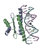 1egwS S: Starting model for refinement |
|---|---|
| Similar structure data |


-Links
 PDBj
PDBj


- Assembly
| Deposited unit | 
| ||||||||
|---|---|---|---|---|---|---|---|---|---|
| 1 |
| ||||||||
| Unit cell |
| ||||||||
| Number of models | 2 |
-Components
| #1: DNA chain | Mass: 4278.815 Da / Num. of mol.: 1 / Source method: obtained synthetically | ||||
|---|---|---|---|---|---|
| #2: DNA chain | Mass: 4278.815 Da / Num. of mol.: 1 / Source method: obtained synthetically | ||||
| #3: Protein | Mass: 10964.712 Da / Num. of mol.: 2 / Fragment: residues 2-91, MADS-box/MEF2S domain Source method: isolated from a genetically manipulated source Source: (gene. exp.) Homo sapiens (human) / Gene: MEF2B OR XMEF2 / Production host:  #4: Protein/peptide | | Mass: 3720.275 Da / Num. of mol.: 1 / Fragment: residues 101-132, Cabin1, MEF2-binding domain Source method: isolated from a genetically manipulated source Source: (gene. exp.) Homo sapiens (human) / Gene: CABIN1 OR KIAA0330 / Production host: #5: Water | ChemComp-HOH / |  Mass: 18.015 Da / Num. of mol.: 228 / Source method: isolated from a natural source / Formula: H2O Mass: 18.015 Da / Num. of mol.: 228 / Source method: isolated from a natural source / Formula: H2O |
-Experimental details
-Experiment
| Experiment | Method: X-RAY DIFFRACTION |
|---|
- Sample preparation
| Crystal | Density Matthews: 2.67 Å3/Da / Density % sol: 53.53 % | |||||||||||||||||||||||||||||||||||||||||||||||||||||||||||||||
|---|---|---|---|---|---|---|---|---|---|---|---|---|---|---|---|---|---|---|---|---|---|---|---|---|---|---|---|---|---|---|---|---|---|---|---|---|---|---|---|---|---|---|---|---|---|---|---|---|---|---|---|---|---|---|---|---|---|---|---|---|---|---|---|---|
| Crystal grow | Temperature: 298 K / Method: vapor diffusion, hanging drop / pH: 7.2 Details: PEG 3500, sodium cholride, BTP, pH 7.2, VAPOR DIFFUSION, HANGING DROP, temperature 298K | |||||||||||||||||||||||||||||||||||||||||||||||||||||||||||||||
| Components of the solutions |
| |||||||||||||||||||||||||||||||||||||||||||||||||||||||||||||||
| Crystal grow | *PLUS Temperature: 17 ℃ / Method: vapor diffusion, hanging drop / PH range low: 6.68 / PH range high: 6.35 | |||||||||||||||||||||||||||||||||||||||||||||||||||||||||||||||
| Components of the solutions | *PLUS
|
-Data collection
| Diffraction | Mean temperature: 101 K |
|---|---|
| Diffraction source | Source: SYNCHROTRON / Site: APS  / Beamline: 14-BM-C / Beamline: 14-BM-C |
| Detector | Type: ADSC QUANTUM 4 / Detector: CCD |
| Radiation | Protocol: SINGLE WAVELENGTH / Monochromatic (M) / Laue (L): M / Scattering type: x-ray |
| Radiation wavelength | Relative weight: 1 |
| Reflection | Resolution: 2.2→30 Å / Num. all: 18733 / Num. obs: 18733 / % possible obs: 90 % / Observed criterion σ(F): 0 / Observed criterion σ(I): 0 / Redundancy: 19.5 % / Biso Wilson estimate: 23.8 Å2 / Rmerge(I) obs: 0.059 / Net I/σ(I): 38 |
| Reflection shell | Resolution: 2.2→2.28 Å / Redundancy: 4.4 % / Rmerge(I) obs: 0.059 / Mean I/σ(I) obs: 4.4 / Num. unique all: 1135 / Rsym value: 0.37 / % possible all: 54 |
| Reflection | *PLUS Highest resolution: 2.2 Å / Lowest resolution: 30 Å / % possible obs: 92.7 % / Num. measured all: 364592 / Rmerge(I) obs: 0.063 |
| Reflection shell | *PLUS Highest resolution: 2.2 Å / Lowest resolution: 2.3 Å / % possible obs: 77.7 % / Rmerge(I) obs: 0.319 |
- Processing
| Software |
| ||||||||||||||||||||||||||||||||||||
|---|---|---|---|---|---|---|---|---|---|---|---|---|---|---|---|---|---|---|---|---|---|---|---|---|---|---|---|---|---|---|---|---|---|---|---|---|---|
| Refinement | Method to determine structure: MOLECULAR REPLACEMENT Starting model: PDB ENTRY 1EGW Resolution: 2.2→28.99 Å / Rfactor Rfree error: 0.006 / Data cutoff high absF: 368477.9 / Data cutoff high rms absF: 368477.9 / Data cutoff low absF: 0 / Isotropic thermal model: RESTRAINED / Cross valid method: THROUGHOUT / σ(F): 0 / σ(I): 0 / Stereochemistry target values: Engh & Huber Details: There are two models represented in this entry (MODEL 1 and MODEL 2) containing 5 chains (ABGCD) each. Molecules ABGCD in MODEL 1 is an alternative conformation to the molecules ABGCD in MODEL 2.
| ||||||||||||||||||||||||||||||||||||
| Solvent computation | Solvent model: FLAT MODEL / Bsol: 41.6301 Å2 / ksol: 0.267209 e/Å3 | ||||||||||||||||||||||||||||||||||||
| Displacement parameters | Biso mean: 53.6 Å2
| ||||||||||||||||||||||||||||||||||||
| Refine analyze |
| ||||||||||||||||||||||||||||||||||||
| Refinement step | Cycle: LAST / Resolution: 2.2→28.99 Å
| ||||||||||||||||||||||||||||||||||||
| Refine LS restraints |
| ||||||||||||||||||||||||||||||||||||
| LS refinement shell | Resolution: 2.2→2.34 Å / Rfactor Rfree error: 0.022 / Total num. of bins used: 6
| ||||||||||||||||||||||||||||||||||||
| Xplor file |
| ||||||||||||||||||||||||||||||||||||
| Refinement | *PLUS Highest resolution: 2.2 Å / Lowest resolution: 30 Å / Num. reflection obs: 18733 / % reflection Rfree: 10 % / Rfactor Rwork: 0.242 | ||||||||||||||||||||||||||||||||||||
| Solvent computation | *PLUS | ||||||||||||||||||||||||||||||||||||
| Displacement parameters | *PLUS | ||||||||||||||||||||||||||||||||||||
| Refine LS restraints | *PLUS
| ||||||||||||||||||||||||||||||||||||
| LS refinement shell | *PLUS Highest resolution: 2.2 Å / Lowest resolution: 2.3 Å / Rfactor Rfree: 0.341 / Rfactor Rwork: 0.313 / Num. reflection Rwork: 1613 |
