 Movie
Movie Controller


+ Open data

- Basic information
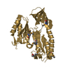
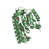
| Entry | Database: PDB / ID: 1egw | ||||||
|---|---|---|---|---|---|---|---|
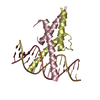
| Title | CRYSTAL STRUCTURE OF MEF2A CORE BOUND TO DNA | ||||||
 Components Components |
| ||||||
 Keywords Keywords | TRANSCRIPTION/DNA / MADS-box transcription factor / DNA-protein complex / TRANSCRIPTION-DNA complex | ||||||
| Function / homology |  Function and homology information Function and homology informationventricular cardiac myofibril assembly / mitochondrion distribution / cardiac conduction / muscle cell development / muscle organ development / Myogenesis / dendrite morphogenesis / histone acetyltransferase binding / positive regulation of cardiac muscle hypertrophy / ERK/MAPK targets ...ventricular cardiac myofibril assembly / mitochondrion distribution / cardiac conduction / muscle cell development / muscle organ development / Myogenesis / dendrite morphogenesis / histone acetyltransferase binding / positive regulation of cardiac muscle hypertrophy / ERK/MAPK targets / SMAD binding / cellular response to calcium ion / positive regulation of D-glucose import across plasma membrane / RNA polymerase II transcription regulatory region sequence-specific DNA binding / histone deacetylase binding / heart development / DNA-binding transcription activator activity, RNA polymerase II-specific / transcription regulator complex / DNA-binding transcription factor binding / sequence-specific DNA binding / RNA polymerase II-specific DNA-binding transcription factor binding / DNA-binding transcription factor activity, RNA polymerase II-specific / cell differentiation / RNA polymerase II cis-regulatory region sequence-specific DNA binding / DNA-binding transcription factor activity / protein heterodimerization activity / apoptotic process / chromatin binding / positive regulation of gene expression / DNA-templated transcription / protein kinase binding / chromatin / negative regulation of transcription by RNA polymerase II / positive regulation of transcription by RNA polymerase II / nucleoplasm / nucleus / cytosol Similarity search - Function | ||||||
| Biological species |  Homo sapiens (human) Homo sapiens (human) | ||||||
| Method |  X-RAY DIFFRACTION / SYNCHROTRON / MOLECULAR REPLACEMENT, heavy atom phases from iodine derivative dataset / Resolution: 1.5 Å X-RAY DIFFRACTION / SYNCHROTRON / MOLECULAR REPLACEMENT, heavy atom phases from iodine derivative dataset / Resolution: 1.5 Å | ||||||
 Authors Authors | Santelli, E. / Richmond, T.J. | ||||||
 Citation Citation | Journal: J.Mol.Biol. / Year: 2000 Title: Crystal structure of MEF2A core bound to DNA at 1.5 A resolution. Authors: Santelli, E. / Richmond, T.J. | ||||||
| History |
|
- Structure visualization
| Structure viewer | Molecule: MolmilJmol/JSmol |
|---|
- Downloads & links
-Download
| PDBx/mmCIF format | 1egw.cif.gz | 153.9 KB | Display | PDBx/mmCIF format |
|---|---|---|---|---|
| PDB format | pdb1egw.ent.gz | 120.8 KB | Display | PDB format |
| PDBx/mmJSON format | 1egw.json.gz | Tree view | PDBx/mmJSON format | |
| Others |  Other downloads Other downloads |
-Validation report
| Arichive directory | https://data.pdbj.org/pub/pdb/validation_reports/eg/1egwftp://data.pdbj.org/pub/pdb/validation_reports/eg/1egw | HTTPS FTP |
|---|
-Related structure data
| Similar structure data |
|---|


-Links
 PDBj
PDBj


- Assembly
| Deposited unit | 
| ||||||||||
|---|---|---|---|---|---|---|---|---|---|---|---|
| 1 | 
| ||||||||||
| 2 | 
| ||||||||||
| Unit cell |
| ||||||||||
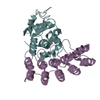
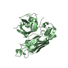
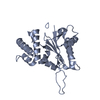
| Details | A protein dimer bound to a double stranded DNA oligonucleotide fully described by the deposited coordinates |
-Components
| #1: DNA chain | Mass: 5200.408 Da / Num. of mol.: 2 / Source method: obtained synthetically / Details: Consensus DNA binding site for MEF2 #2: DNA chain | Mass: 5209.422 Da / Num. of mol.: 2 / Source method: obtained synthetically / Details: Consensus DNA binding site for MEF2 #3: Protein | Mass: 9155.646 Da / Num. of mol.: 4 / Fragment: N-TERMINUS, RESIDUES 2-78 Source method: isolated from a genetically manipulated source Source: (gene. exp.) Homo sapiens (human) / Organ: HEART, SKELETAL MUSCLE / Production host:  #4: Water | ChemComp-HOH / |  Mass: 18.015 Da / Num. of mol.: 656 / Source method: isolated from a natural source / Formula: H2O Mass: 18.015 Da / Num. of mol.: 656 / Source method: isolated from a natural source / Formula: H2O |
|---|
-Experimental details
-Experiment
| Experiment | Method: X-RAY DIFFRACTION / Number of used crystals: 3 |
|---|
- Sample preparation
| Crystal | Density Matthews: 2.5 Å3/Da / Density % sol: 50.84 % | ||||||||||||||||||||||||||||||||||||||||||||||||
|---|---|---|---|---|---|---|---|---|---|---|---|---|---|---|---|---|---|---|---|---|---|---|---|---|---|---|---|---|---|---|---|---|---|---|---|---|---|---|---|---|---|---|---|---|---|---|---|---|---|
| Crystal grow | Temperature: 295 K / Method: vapor diffusion, hanging drop Details: PEG 6000, NaCl, Ba(NO3)2, sodium Acetate, Bis-tris buffer, DTT, VAPOR DIFFUSION, HANGING DROP, temperature 295K PH range: 5.8-6.3 | ||||||||||||||||||||||||||||||||||||||||||||||||
| Components of the solutions |
| ||||||||||||||||||||||||||||||||||||||||||||||||
| Crystal grow | *PLUS Temperature: 22 ℃ | ||||||||||||||||||||||||||||||||||||||||||||||||
| Components of the solutions | *PLUS
|
-Data collection
| Diffraction | Mean temperature: 110 K |
|---|---|
| Diffraction source | Source: SYNCHROTRON / Site: ESRF  / Type: ESRF / Wavelength: 0.932 / Type: ESRF / Wavelength: 0.932 |
| Detector | Type: MARRESEARCH / Detector: IMAGE PLATE / Date: May 18, 1999 |
| Radiation | Protocol: SINGLE WAVELENGTH / Monochromatic (M) / Laue (L): M / Scattering type: x-ray |
| Radiation wavelength | Wavelength: 0.932 Å / Relative weight: 1 |
| Reflection | Resolution: 1.5→30 Å / Num. obs: 87264 / % possible obs: 96 % / Observed criterion σ(F): 0 / Observed criterion σ(I): 0 / Redundancy: 2.6 % / Biso Wilson estimate: 14.7 Å2 / Rmerge(I) obs: 0.075 / Net I/σ(I): 12.8 |
| Reflection shell | Resolution: 1.5→1.55 Å / Redundancy: 1.9 % / Rmerge(I) obs: 0.104 / % possible all: 92 |
| Reflection shell | *PLUS % possible obs: 92 % |
- Processing
| Software |
| ||||||||||||||||||||||||||||||||||||||||
|---|---|---|---|---|---|---|---|---|---|---|---|---|---|---|---|---|---|---|---|---|---|---|---|---|---|---|---|---|---|---|---|---|---|---|---|---|---|---|---|---|---|
| Refinement | Method to determine structure: MOLECULAR REPLACEMENT, heavy atom phases from iodine derivative dataset Resolution: 1.5→20 Å / Rfactor Rfree error: 0.002 / Data cutoff high absF: 1092727.38 / Data cutoff low absF: 0 / Isotropic thermal model: RESTRAINED / Cross valid method: THROUGHOUT / σ(F): 0 / σ(I): 0 / Stereochemistry target values: Engh & Huber Details: refined also with REFMAC by Murshudov, Vagin, Dodson
| ||||||||||||||||||||||||||||||||||||||||
| Solvent computation | Solvent model: FLAT MODEL / Bsol: 51.92 Å2 / ksol: 0.316 e/Å3 | ||||||||||||||||||||||||||||||||||||||||
| Displacement parameters | Biso mean: 24.1 Å2
| ||||||||||||||||||||||||||||||||||||||||
| Refine analyze |
| ||||||||||||||||||||||||||||||||||||||||
| Refinement step | Cycle: LAST / Resolution: 1.5→20 Å
| ||||||||||||||||||||||||||||||||||||||||
| Refine LS restraints |
| ||||||||||||||||||||||||||||||||||||||||
| LS refinement shell | Resolution: 1.5→1.57 Å / Rfactor Rfree error: 0.009 / Total num. of bins used: 8
| ||||||||||||||||||||||||||||||||||||||||
| Xplor file |
| ||||||||||||||||||||||||||||||||||||||||
| Software | *PLUS Name: CNS / Version: 0.9 / Classification: refinement | ||||||||||||||||||||||||||||||||||||||||
| Refinement | *PLUS Lowest resolution: 20 Å / σ(F): 0 / % reflection Rfree: 10 % / Rfactor obs: 0.206 | ||||||||||||||||||||||||||||||||||||||||
| Solvent computation | *PLUS | ||||||||||||||||||||||||||||||||||||||||
| Displacement parameters | *PLUS Biso mean: 24.1 Å2 | ||||||||||||||||||||||||||||||||||||||||
| Refine LS restraints | *PLUS
| ||||||||||||||||||||||||||||||||||||||||
| LS refinement shell | *PLUS Rfactor Rfree: 0.286 / % reflection Rfree: 10.2 % / Rfactor Rwork: 0.257 / Rfactor obs: 0.257 |
