 Movie
Movie Controller

[English] 日本語
 Yorodumi
- PDB-2ql2: Crystal Structure of the basic-helix-loop-helix domains of the he... -
+ Open data

- Basic information
| Entry | Database: PDB / ID: 2ql2 | ||||||
|---|---|---|---|---|---|---|---|
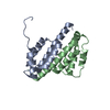
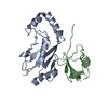
| Title | Crystal Structure of the basic-helix-loop-helix domains of the heterodimer E47/NeuroD1 bound to DNA | ||||||
 Components Components |
| ||||||
 Keywords Keywords | transcription/DNA / basic-helix-loop-helix / protein-DNA complex / heterodimer / DNA-binding / Activator / Developmental protein / Differentiation / Neurogenesis / Nucleus / Phosphorylation / Transcription / Transcription regulation / Cytoplasm / Phosphoprotein / transcription-DNA COMPLEX | ||||||
| Function / homology |  Function and homology information Function and homology informationpancreatic A cell fate commitment / pancreatic PP cell fate commitment / regulation of intestinal epithelial structure maintenance / enteroendocrine cell differentiation / vitamin D response element binding / negative regulation of type B pancreatic cell apoptotic process / amacrine cell differentiation / bHLH transcription factor binding / embryonic organ morphogenesis / cell development ...pancreatic A cell fate commitment / pancreatic PP cell fate commitment / regulation of intestinal epithelial structure maintenance / enteroendocrine cell differentiation / vitamin D response element binding / negative regulation of type B pancreatic cell apoptotic process / amacrine cell differentiation / bHLH transcription factor binding / embryonic organ morphogenesis / cell development / natural killer cell differentiation / RUNX1 regulates transcription of genes involved in differentiation of HSCs / lymphocyte differentiation / hindbrain development / endocrine pancreas development / sensory organ development / Peyer's patch development / immunoglobulin V(D)J recombination / Myogenesis / dentate gyrus development / negative regulation of receptor signaling pathway via JAK-STAT / anterior/posterior pattern specification / camera-type eye development / signal transduction involved in regulation of gene expression / mitogen-activated protein kinase kinase kinase binding / insulin secretion / regulation of neuron differentiation / nucleocytoplasmic transport / cellular response to glucocorticoid stimulus / B cell lineage commitment / axon development / inner ear development / E-box binding / cell fate commitment / response to glucose / cis-regulatory region sequence-specific DNA binding / regulation of G1/S transition of mitotic cell cycle / positive regulation of B cell proliferation / gastrulation / positive regulation of neuron differentiation / positive regulation of cell cycle / cerebellum development / PDZ domain binding / cellular response to glucose stimulus / positive regulation of cell differentiation / euchromatin / DNA-binding transcription repressor activity, RNA polymerase II-specific / RNA polymerase II transcription regulator complex / positive regulation of insulin secretion / sequence-specific double-stranded DNA binding / T cell differentiation in thymus / nucleosome / glucose homeostasis / nervous system development / double-stranded DNA binding / DNA-binding transcription activator activity, RNA polymerase II-specific / transcription regulator complex / response to lipopolysaccharide / DNA-binding transcription factor binding / sequence-specific DNA binding / RNA polymerase II-specific DNA-binding transcription factor binding / DNA-binding transcription factor activity, RNA polymerase II-specific / protein stabilization / response to xenobiotic stimulus / RNA polymerase II cis-regulatory region sequence-specific DNA binding / positive regulation of apoptotic process / chromatin remodeling / DNA-binding transcription factor activity / protein heterodimerization activity / chromatin binding / positive regulation of gene expression / regulation of transcription by RNA polymerase II / regulation of DNA-templated transcription / positive regulation of DNA-templated transcription / chromatin / negative regulation of transcription by RNA polymerase II / DNA-templated transcription / protein homodimerization activity / positive regulation of transcription by RNA polymerase II / protein-containing complex / DNA binding / nucleoplasm / identical protein binding / nucleus / cytosol / cytoplasm Similarity search - Function | ||||||
| Biological species |  | ||||||
| Method |  X-RAY DIFFRACTION / SYNCHROTRON / MOLECULAR REPLACEMENT / Resolution: 2.5 Å X-RAY DIFFRACTION / SYNCHROTRON / MOLECULAR REPLACEMENT / Resolution: 2.5 Å | ||||||
 Authors Authors | Rose, R.B. / Longo, A. / Guanga, G.P. | ||||||
 Citation Citation | Journal: Biochemistry / Year: 2008 Title: Crystal structure of E47-NeuroD1/beta2 bHLH domain-DNA complex: heterodimer selectivity and DNA recognition. Authors: Longo, A. / Guanga, G.P. / Rose, R.B. | ||||||
| History |
|
- Structure visualization
| Structure viewer | Molecule: MolmilJmol/JSmol |
|---|
- Downloads & links
-Download
| PDBx/mmCIF format | 2ql2.cif.gz | 94.5 KB | Display | PDBx/mmCIF format |
|---|---|---|---|---|
| PDB format | pdb2ql2.ent.gz | 67.1 KB | Display | PDB format |
| PDBx/mmJSON format | 2ql2.json.gz | Tree view | PDBx/mmJSON format | |
| Others |  Other downloads Other downloads |
-Validation report
| Arichive directory | https://data.pdbj.org/pub/pdb/validation_reports/ql/2ql2ftp://data.pdbj.org/pub/pdb/validation_reports/ql/2ql2 | HTTPS FTP |
|---|
-Related structure data
| Related structure data | 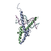 1mdyS S: Starting model for refinement |
|---|---|
| Similar structure data |


-Links
 PDBj
PDBj


- Assembly
| Deposited unit | 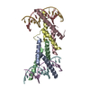
| ||||||||
|---|---|---|---|---|---|---|---|---|---|
| 1 | 
| ||||||||
| 2 | 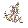
| ||||||||
| 3 |
| ||||||||
| Unit cell |
| ||||||||
| Details | The biological assembly is a heterodimer of E47 and NeuroD1 bound to a DNA duplex. There are 2 of these complexes in the asymmetric unit. |
-Components
| #1: Protein | Mass: 7141.364 Da / Num. of mol.: 2 / Fragment: basic-helix-loop-helix domain Source method: isolated from a genetically manipulated source Details: pancreatic beta cells / Source: (gene. exp.)  #2: Protein | Mass: 6986.231 Da / Num. of mol.: 2 / Fragment: basic helix-loop-helix domain Source method: isolated from a genetically manipulated source Details: pancreatic beta cells / Source: (gene. exp.) #3: DNA chain | Mass: 4865.152 Da / Num. of mol.: 2 / Source method: obtained synthetically / Details: synthesized oligonucleotide #4: DNA chain | Mass: 4932.218 Da / Num. of mol.: 2 / Source method: obtained synthetically / Details: synthesized oligonucleotide #5: Water | ChemComp-HOH / |  Mass: 18.015 Da / Num. of mol.: 41 / Source method: isolated from a natural source / Formula: H2O Mass: 18.015 Da / Num. of mol.: 41 / Source method: isolated from a natural source / Formula: H2OSequence details | THE PROTEIN IN CHAINS A AND C MATCH THE UNP SEQUENCE DATABASE REFERENCE P15806(ISOFORM E47; P15806- ...THE PROTEIN IN CHAINS A AND C MATCH THE UNP SEQUENCE DATABASE REFERENCE P15806(ISOFORM E47; P15806-2), RESIDUES 544-603 | |
|---|
-Experimental details
-Experiment
| Experiment | Method: X-RAY DIFFRACTION / Number of used crystals: 1 |
|---|
- Sample preparation
| Crystal | Density Matthews: 2.79 Å3/Da / Density % sol: 55.93 % | ||||||||||||||||||||
|---|---|---|---|---|---|---|---|---|---|---|---|---|---|---|---|---|---|---|---|---|---|
| Crystal grow | Temperature: 291 K / Method: evaporation / pH: 7.6 Details: 0.1 mM Imidazole, 0.2 M ammonium citrate dibasic, and 22 % PEG 4000, pH 7.6, EVAPORATION, temperature 291K | ||||||||||||||||||||
| Components of the solutions |
|
-Data collection
| Diffraction | Mean temperature: 100 K |
|---|---|
| Diffraction source | Source: SYNCHROTRON / Site: APS  / Beamline: 22-BM / Wavelength: 1 Å / Beamline: 22-BM / Wavelength: 1 Å |
| Detector | Type: MAR scanner 345 mm plate / Detector: IMAGE PLATE / Date: Dec 1, 2006 Details: sagittal focusing crystal and vertical focusing double mirror |
| Radiation | Monochromator: SI (111) double crystal / Protocol: SINGLE WAVELENGTH / Monochromatic (M) / Laue (L): M / Scattering type: x-ray |
| Radiation wavelength | Wavelength: 1 Å / Relative weight: 1 |
| Reflection | Resolution: 2.5→169.546 Å / Num. obs: 18832 / % possible obs: 97.7 % / Observed criterion σ(I): 1 / Redundancy: 7.2 % / Biso Wilson estimate: 76 Å2 / Rmerge(I) obs: 0.076 / Rsym value: 0.076 / Net I/σ(I): 5.7 |
| Reflection shell | Resolution: 2.5→2.64 Å / Redundancy: 7.1 % / Rmerge(I) obs: 0.77 / Mean I/σ(I) obs: 1 / Num. measured all: 18434 / Num. unique all: 2594 / Rsym value: 0.77 / % possible all: 94.8 |
- Processing
| Software |
| ||||||||||||||||||||||||||||||||
|---|---|---|---|---|---|---|---|---|---|---|---|---|---|---|---|---|---|---|---|---|---|---|---|---|---|---|---|---|---|---|---|---|---|
| Refinement | Method to determine structure: MOLECULAR REPLACEMENT Starting model: pdb entry 1MDY Resolution: 2.5→55.8 Å / Isotropic thermal model: isotropic / Cross valid method: THROUGHOUT / σ(F): 0 / Stereochemistry target values: Engh & Huber Details: optimal B-restraints weight (rweight) in CNS was 0.0198
| ||||||||||||||||||||||||||||||||
| Solvent computation | Bsol: 83.749 Å2 | ||||||||||||||||||||||||||||||||
| Displacement parameters | Biso mean: 56.998 Å2
| ||||||||||||||||||||||||||||||||
| Refine analyze |
| ||||||||||||||||||||||||||||||||
| Refinement step | Cycle: LAST / Resolution: 2.5→55.8 Å
| ||||||||||||||||||||||||||||||||
| Refine LS restraints |
| ||||||||||||||||||||||||||||||||
| LS refinement shell | Resolution: 2.5→2.59 Å
| ||||||||||||||||||||||||||||||||
| Xplor file |
|
