 Movie
Movie Controller

[English] 日本語
 Yorodumi
- PDB-1mdy: CRYSTAL STRUCTURE OF MYOD BHLH DOMAIN BOUND TO DNA: PERSPECTIVES ... -
+ Open data

- Basic information
| Entry | Database: PDB / ID: 1mdy | ||||||
|---|---|---|---|---|---|---|---|
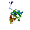
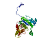
| Title | CRYSTAL STRUCTURE OF MYOD BHLH DOMAIN BOUND TO DNA: PERSPECTIVES ON DNA RECOGNITION AND IMPLICATIONS FOR TRANSCRIPTIONAL ACTIVATION | ||||||
 Components Components |
| ||||||
 Keywords Keywords | TRANSCRIPTION/DNA / PROTEIN-DNA COMPLEX / TRANSCRIPTION-DNA COMPLEX | ||||||
| Function / homology |  Function and homology information Function and homology informationmyoblast fate determination / myotube differentiation involved in skeletal muscle regeneration / skeletal muscle fiber adaptation / negative regulation of myoblast proliferation / positive regulation of snRNA transcription by RNA polymerase II / positive regulation of skeletal muscle tissue regeneration / positive regulation of skeletal muscle fiber development / myotube differentiation / myotube cell development / bHLH transcription factor binding ...myoblast fate determination / myotube differentiation involved in skeletal muscle regeneration / skeletal muscle fiber adaptation / negative regulation of myoblast proliferation / positive regulation of snRNA transcription by RNA polymerase II / positive regulation of skeletal muscle tissue regeneration / positive regulation of skeletal muscle fiber development / myotube differentiation / myotube cell development / bHLH transcription factor binding / muscle cell differentiation / Myogenesis / cardiac muscle cell differentiation / skeletal muscle tissue regeneration / myoblast fusion / myoblast differentiation / cellular response to oxygen levels / positive regulation of myoblast fusion / cellular response to glucocorticoid stimulus / ATPase complex / positive regulation of muscle cell differentiation / muscle organ development / DNA-binding transcription activator activity / regulation of alternative mRNA splicing, via spliceosome / regulation of RNA splicing / E-box binding / skeletal muscle cell differentiation / myofibril / protein unfolding / positive regulation of myoblast differentiation / skeletal muscle tissue development / cis-regulatory region sequence-specific DNA binding / skeletal muscle fiber development / striated muscle cell differentiation / nuclear receptor binding / cellular response to starvation / RNA polymerase II transcription regulatory region sequence-specific DNA binding / cellular response to estradiol stimulus / promoter-specific chromatin binding / euchromatin / chromatin DNA binding / cellular response to tumor necrosis factor / sequence-specific double-stranded DNA binding / regulation of gene expression / DNA-binding transcription activator activity, RNA polymerase II-specific / transcription regulator complex / sequence-specific DNA binding / RNA polymerase II-specific DNA-binding transcription factor binding / DNA-binding transcription factor activity, RNA polymerase II-specific / RNA polymerase II cis-regulatory region sequence-specific DNA binding / DNA-binding transcription factor activity / chromatin binding / ubiquitin protein ligase binding / regulation of transcription by RNA polymerase II / positive regulation of DNA-templated transcription / chromatin / enzyme binding / DNA-templated transcription / protein homodimerization activity / positive regulation of transcription by RNA polymerase II / nucleoplasm / nucleus / cytoplasm Similarity search - Function | ||||||
| Biological species |  | ||||||
| Method |  X-RAY DIFFRACTION / Resolution: 2.8 Å X-RAY DIFFRACTION / Resolution: 2.8 Å | ||||||
 Authors Authors | Ma, P.C.M. / Rould, M.A. / Weintraub, H. / Pabo, C.O. | ||||||
 Citation Citation | Journal: Cell(Cambridge,Mass.) / Year: 1994 Title: Crystal structure of MyoD bHLH domain-DNA complex: perspectives on DNA recognition and implications for transcriptional activation. Authors: Ma, P.C. / Rould, M.A. / Weintraub, H. / Pabo, C.O. | ||||||
| History |
|
- Structure visualization
| Structure viewer | Molecule: MolmilJmol/JSmol |
|---|
- Downloads & links
-Download
| PDBx/mmCIF format | 1mdy.cif.gz | 94.1 KB | Display | PDBx/mmCIF format |
|---|---|---|---|---|
| PDB format | pdb1mdy.ent.gz | 69.8 KB | Display | PDB format |
| PDBx/mmJSON format | 1mdy.json.gz | Tree view | PDBx/mmJSON format | |
| Others |  Other downloads Other downloads |
-Validation report
| Arichive directory | https://data.pdbj.org/pub/pdb/validation_reports/md/1mdyftp://data.pdbj.org/pub/pdb/validation_reports/md/1mdy | HTTPS FTP |
|---|
-Related structure data
| Similar structure data |
|---|


-Links
 PDBj
PDBj


- Assembly
| Deposited unit | 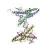
| ||||||||||||
|---|---|---|---|---|---|---|---|---|---|---|---|---|---|
| 1 | 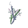
| ||||||||||||
| 2 | 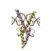
| ||||||||||||
| Unit cell |
| ||||||||||||
| Noncrystallographic symmetry (NCS) | NCS oper:
| ||||||||||||
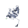
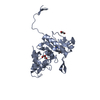
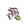
| Details | THE ASYMMETRIC UNIT CONTAINS FOUR MONOMERS OF MYOD TOGETHER WITH TWO DOUBLE-STRANDED 14 BASE PAIR OLIGONUCLEOTIDES. THERE ARE, THUS, TWO HOMODIMERS OF MYOD BOUND TO TWO DNA SITES IN THE ASYMMETRIC UNIT. THE DEPOSITORS HAVE INCLUDED RESIDUES 105 - 166 OF ALL FOUR OF THE MYOD MONOMERS IN THEIR MODEL. RESIDUES 1 - 3 AND 102 - 104 ARE ALSO INCLUDED IN ONE OUT OF THE FOUR MONOMERS, WHERE THESE RESIDUES ARE INVOLVED IN CRYSTAL PACKING CONTACTS. THE TRANSFORMATION PRESENTED ON *MTRIX 1* RECORDS BELOW WILL YIELD APPROXIMATE COORDINATES FOR CHAIN *B* WHEN APPLIED TO CHAIN *A*. THE TRANSFORMATION PRESENTED ON *MTRIX 2* RECORDS BELOW WILL YIELD APPROXIMATE COORDINATES FOR CHAIN *D* WHEN APPLIED TO CHAIN *C*. |
-Components
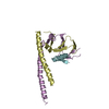
| #1: DNA chain | Mass: 4279.804 Da / Num. of mol.: 4 / Source method: obtained synthetically #2: Protein | | Mass: 8058.361 Da / Num. of mol.: 1 / Source method: isolated from a natural source / Source: (natural) #3: Protein | Mass: 7269.341 Da / Num. of mol.: 3 / Source method: isolated from a natural source / Source: (natural) #4: Water | ChemComp-HOH / |  Mass: 18.015 Da / Num. of mol.: 25 / Source method: isolated from a natural source / Formula: H2O Mass: 18.015 Da / Num. of mol.: 25 / Source method: isolated from a natural source / Formula: H2OCompound details | DNA SYNTHETIC OLIGONUCLEOTIDE OF 14 BASE PAIRS, CONTAINING THE OPTIMIZED DNA BINDING SITE FOR THE ...DNA SYNTHETIC OLIGONUCLE | Sequence details | THE PROTEIN RESIDUES ARE NUMBERED ACCORDING TO THE NATIVE SCHEME FOR MOUSE MYOD PROTEIN. THERE ARE ...THE PROTEIN RESIDUES ARE NUMBERED ACCORDING TO THE NATIVE SCHEME FOR MOUSE MYOD PROTEIN. THERE ARE FOUR SEPARATE MYOD MONOMERS IN THE ASYMMETRIC | |
|---|
-Experimental details
-Experiment
| Experiment | Method: X-RAY DIFFRACTION |
|---|
- Sample preparation
| Crystal | Density Matthews: 2.48 Å3/Da / Density % sol: 50.48 % | ||||||||||||||||||||||||||||||
|---|---|---|---|---|---|---|---|---|---|---|---|---|---|---|---|---|---|---|---|---|---|---|---|---|---|---|---|---|---|---|---|
| Crystal | *PLUS Density % sol: 55 % | ||||||||||||||||||||||||||||||
| Crystal grow | *PLUS Temperature: 22 ℃ / pH: 8.5 / Method: vapor diffusion, hanging drop | ||||||||||||||||||||||||||||||
| Components of the solutions | *PLUS
|
-Data collection
| Radiation | Protocol: SINGLE WAVELENGTH / Monochromatic (M) / Laue (L): M / Scattering type: x-ray |
|---|---|
| Radiation wavelength | Relative weight: 1 |
- Processing
| Software |
| ||||||||||||
|---|---|---|---|---|---|---|---|---|---|---|---|---|---|
| Refinement | Rfactor Rfree: 0.33 / Rfactor Rwork: 0.253 / Rfactor obs: 0.253 / Highest resolution: 2.8 Å | ||||||||||||
| Refinement step | Cycle: LAST / Highest resolution: 2.8 Å
| ||||||||||||
| Refinement | *PLUS Highest resolution: 2.8 Å / Lowest resolution: 20 Å / Num. reflection all: 10585 / Num. reflection obs: 8963 / σ(I): 2 / Rfactor obs: 0.224 / Rfactor Rfree: 0.33 | ||||||||||||
| Solvent computation | *PLUS | ||||||||||||
| Displacement parameters | *PLUS |
