 Movie
Movie Controller

[English] 日本語
 Yorodumi
- PDB-1jrg: Crystal Structure of the R3 form of Pectate Lyase A, Erwinia chry... -
+ Open data

- Basic information
| Entry | Database: PDB / ID: 1jrg | ||||||
|---|---|---|---|---|---|---|---|
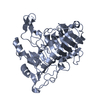
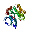
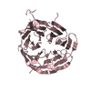
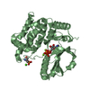
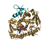
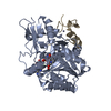
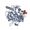
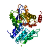
| Title | Crystal Structure of the R3 form of Pectate Lyase A, Erwinia chrysanthemi | ||||||
 Components Components | Pectate lyase | ||||||
 Keywords Keywords | LYASE / parallel beta helix beta elimination | ||||||
| Function / homology |  Function and homology information Function and homology informationpectate lyase / pectate lyase activity / pectin catabolic process / extracellular region / metal ion binding Similarity search - Function | ||||||
| Biological species |  Erwinia chrysanthemi (bacteria) Erwinia chrysanthemi (bacteria) | ||||||
| Method |  X-RAY DIFFRACTION / MOLECULAR REPLACEMENT / Resolution: 2.1 Å X-RAY DIFFRACTION / MOLECULAR REPLACEMENT / Resolution: 2.1 Å | ||||||
 Authors Authors | Thomas, L.M. / Doan, C. / Oliver, R.L. / Yoder, M.D. | ||||||
 Citation Citation | Journal: Acta Crystallogr.,Sect.D / Year: 2002 Title: Structure of pectate lyase A: comparison to other isoforms. Authors: Thomas, L.M. / Doan, C.N. / Oliver, R.L. / Yoder, M.D. | ||||||
| History |
|
- Structure visualization
| Structure viewer | Molecule: MolmilJmol/JSmol |
|---|
- Downloads & links
-Download
| PDBx/mmCIF format | 1jrg.cif.gz | 148.6 KB | Display | PDBx/mmCIF format |
|---|---|---|---|---|
| PDB format | pdb1jrg.ent.gz | 116.9 KB | Display | PDB format |
| PDBx/mmJSON format | 1jrg.json.gz | Tree view | PDBx/mmJSON format | |
| Others |  Other downloads Other downloads |
-Validation report
| Arichive directory | https://data.pdbj.org/pub/pdb/validation_reports/jr/1jrgftp://data.pdbj.org/pub/pdb/validation_reports/jr/1jrg | HTTPS FTP |
|---|
-Related structure data


-Links
 PDBj
PDBj- Assembly
| Deposited unit | 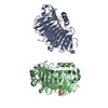
| ||||||||
|---|---|---|---|---|---|---|---|---|---|
| 1 | 
| ||||||||
| 2 | 
| ||||||||
| Unit cell |
|
-Components
| #1: Protein | Mass: 38794.609 Da / Num. of mol.: 2 / Fragment: Pectate lyase Source method: isolated from a genetically manipulated source Source: (gene. exp.) Erwinia chrysanthemi (bacteria) / Genus: Dickeya / Strain: EC16 / Gene: PELA / Plasmid: pPEL812 / Production host: References: UniProt: P29155, UniProt: P0C1A2*PLUS, pectate lyase #2: Chemical | ChemComp-SO4 /   Mass: 96.063 Da / Num. of mol.: 4 / Source method: obtained synthetically / Formula: SO4 Mass: 96.063 Da / Num. of mol.: 4 / Source method: obtained synthetically / Formula: SO4#3: Water | ChemComp-HOH / |  Mass: 18.015 Da / Num. of mol.: 326 / Source method: isolated from a natural source / Formula: H2O Mass: 18.015 Da / Num. of mol.: 326 / Source method: isolated from a natural source / Formula: H2OHas protein modification | Y | |
|---|
-Experimental details
-Experiment
| Experiment | Method: X-RAY DIFFRACTION / Number of used crystals: 1 |
|---|
- Sample preparation
| Crystal | Density Matthews: 2.59 Å3/Da / Density % sol: 52.49 % | ||||||||||||||||||||||||||||||||||||||||||
|---|---|---|---|---|---|---|---|---|---|---|---|---|---|---|---|---|---|---|---|---|---|---|---|---|---|---|---|---|---|---|---|---|---|---|---|---|---|---|---|---|---|---|---|
| Crystal grow | Temperature: 295 K / Method: vapor diffusion, hanging drop / pH: 7 Details: PEG 8K, PEG1K, HEPES, pH 7.0, VAPOR DIFFUSION, HANGING DROP, temperature 295K | ||||||||||||||||||||||||||||||||||||||||||
| Crystal grow | *PLUS Method: vapor diffusion, sitting drop | ||||||||||||||||||||||||||||||||||||||||||
| Components of the solutions | *PLUS
|
-Data collection
| Diffraction | Mean temperature: 153 K |
|---|---|
| Diffraction source | Source: ROTATING ANODE / Type: RIGAKU RU200 / Wavelength: 1.5418 Å |
| Detector | Type: MARRESEARCH / Detector: IMAGE PLATE / Date: Apr 12, 2000 |
| Radiation | Monochromator: graphite / Protocol: SINGLE WAVELENGTH / Monochromatic (M) / Laue (L): M / Scattering type: x-ray |
| Radiation wavelength | Wavelength: 1.5418 Å / Relative weight: 1 |
| Reflection | Resolution: 2.1→50 Å / Num. all: 45453 / Num. obs: 41984 / % possible obs: 92 % / Observed criterion σ(F): 0 / Observed criterion σ(I): 0 / Rsym value: 0.077 / Net I/σ(I): 12 |
| Reflection shell | Resolution: 2.1→2.18 Å / Mean I/σ(I) obs: 3.5 / Rsym value: 0.31 / % possible all: 99 |
| Reflection | *PLUS Num. obs: 44138 / % possible obs: 98.3 % / Num. measured all: 111710 / Rmerge(I) obs: 0.077 |
| Reflection shell | *PLUS % possible obs: 99.8 % / Rmerge(I) obs: 0.31 |
- Processing
| Software |
| |||||||||||||||||||||||||
|---|---|---|---|---|---|---|---|---|---|---|---|---|---|---|---|---|---|---|---|---|---|---|---|---|---|---|
| Refinement | Method to determine structure: MOLECULAR REPLACEMENT Starting model: PelE search model comprised of residues 30-61, 70-100, 105-251, 258-280, 293-314, and 323-332. (Coordinates provided by F. Jurnak) Resolution: 2.1→50 Å / Cross valid method: THROUGHOUT / σ(F): 0 / σ(I): 0 / Stereochemistry target values: Engh & Huber
| |||||||||||||||||||||||||
| Displacement parameters |
| |||||||||||||||||||||||||
| Refinement step | Cycle: LAST / Resolution: 2.1→50 Å
| |||||||||||||||||||||||||
| Refine LS restraints |
| |||||||||||||||||||||||||
| LS refinement shell | Resolution: 2.1→2.2 Å / Total num. of bins used: 8 /
| |||||||||||||||||||||||||
| Xplor file |
| |||||||||||||||||||||||||
| Refinement | *PLUS Rfactor obs: 0.16 / Rfactor Rfree: 0.2109 / Rfactor Rwork: 0.1596 | |||||||||||||||||||||||||
| Solvent computation | *PLUS | |||||||||||||||||||||||||
| Displacement parameters | *PLUS | |||||||||||||||||||||||||
| Refine LS restraints | *PLUS
| |||||||||||||||||||||||||
| LS refinement shell | *PLUS Rfactor Rfree: 0.261 |
