 Movie
Movie Controller


+ Open data

- Basic information
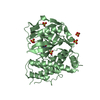
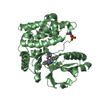
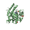
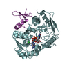
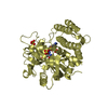
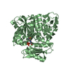
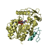
| Entry | Database: PDB / ID: 4pjv | ||||||
|---|---|---|---|---|---|---|---|
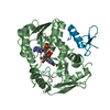
| Title | Structure of PARP2 catalytic domain bound to inhibitor BMN 673 | ||||||
 Components Components | Poly [ADP-ribose] polymerase 2 | ||||||
 Keywords Keywords | TRANSFERASE/TRANSFERASE INHIBITOR / PARP2 / Inhibitor / Complex / TRANSFERASE-TRANSFERASE INHIBITOR complex | ||||||
| Function / homology |  Function and homology information Function and homology informationresponse to oxygen-glucose deprivation / poly-ADP-D-ribose binding / positive regulation of cell growth involved in cardiac muscle cell development / NAD+-protein-serine ADP-ribosyltransferase activity / NAD DNA ADP-ribosyltransferase activity / DNA ADP-ribosylation / poly-ADP-D-ribose modification-dependent protein binding / HDR through MMEJ (alt-NHEJ) / hippocampal neuron apoptotic process / NAD+ ADP-ribosyltransferase ...response to oxygen-glucose deprivation / poly-ADP-D-ribose binding / positive regulation of cell growth involved in cardiac muscle cell development / NAD+-protein-serine ADP-ribosyltransferase activity / NAD DNA ADP-ribosyltransferase activity / DNA ADP-ribosylation / poly-ADP-D-ribose modification-dependent protein binding / HDR through MMEJ (alt-NHEJ) / hippocampal neuron apoptotic process / NAD+ ADP-ribosyltransferase / protein auto-ADP-ribosylation / NAD+-protein-aspartate ADP-ribosyltransferase activity / protein poly-ADP-ribosylation / NAD+-protein-glutamate ADP-ribosyltransferase activity / NAD+-protein mono-ADP-ribosyltransferase activity / DNA repair-dependent chromatin remodeling / decidualization / Transferases; Glycosyltransferases; Pentosyltransferases / POLB-Dependent Long Patch Base Excision Repair / NAD+ poly-ADP-ribosyltransferase activity / nucleosome binding / site of DNA damage / extrinsic apoptotic signaling pathway / nucleotidyltransferase activity / base-excision repair / DNA Damage Recognition in GG-NER / Dual Incision in GG-NER / Formation of Incision Complex in GG-NER / double-strand break repair / damaged DNA binding / DNA repair / chromatin binding / DNA damage response / nucleolus / nucleoplasm / nucleus / cytosol Similarity search - Function | ||||||
| Biological species |  Homo sapiens (human) Homo sapiens (human) | ||||||
| Method |  X-RAY DIFFRACTION / SYNCHROTRON / MOLECULAR REPLACEMENT / Resolution: 2.5 Å X-RAY DIFFRACTION / SYNCHROTRON / MOLECULAR REPLACEMENT / Resolution: 2.5 Å | ||||||
 Authors Authors | Aoyagi-Scharber, M. / Gardberg, A.S. / Edwards, T.L. | ||||||
 Citation Citation | Journal: Acta Crystallogr.,Sect.F / Year: 2014 Title: Structural basis for the inhibition of poly(ADP-ribose) polymerases 1 and 2 by BMN 673, a potent inhibitor derived from dihydropyridophthalazinone. Authors: Aoyagi-Scharber, M. / Gardberg, A.S. / Yip, B.K. / Wang, B. / Shen, Y. / Fitzpatrick, P.A. | ||||||
| History |
|
- Structure visualization
| Structure viewer | Molecule: MolmilJmol/JSmol |
|---|
- Downloads & links
-Download
| PDBx/mmCIF format | 4pjv.cif.gz | 270.8 KB | Display | PDBx/mmCIF format |
|---|---|---|---|---|
| PDB format | pdb4pjv.ent.gz | 216.3 KB | Display | PDB format |
| PDBx/mmJSON format | 4pjv.json.gz | Tree view | PDBx/mmJSON format | |
| Others |  Other downloads Other downloads |
-Validation report
| Arichive directory | https://data.pdbj.org/pub/pdb/validation_reports/pj/4pjvftp://data.pdbj.org/pub/pdb/validation_reports/pj/4pjv | HTTPS FTP |
|---|
-Related structure data
| Related structure data |  4pjtC  3kczS C: citing same article ( S: Starting model for refinement |
|---|---|
| Similar structure data |

-Links
 PDBj
PDBj

- Assembly
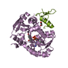
| Deposited unit | 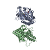
| ||||||||||||||||||
|---|---|---|---|---|---|---|---|---|---|---|---|---|---|---|---|---|---|---|---|
| 1 | 
| ||||||||||||||||||
| 2 | 
| ||||||||||||||||||
| Unit cell |
| ||||||||||||||||||
| Noncrystallographic symmetry (NCS) | NCS domain:
NCS domain segments: Component-ID: 1 / Ens-ID: 1 / Beg auth comp-ID: MET / Beg label comp-ID: MET / End auth comp-ID: PHE / End label comp-ID: PHE / Refine code: 4 / Auth seq-ID: 234 - 579 / Label seq-ID: 23 - 368
|
-Components
| #1: Protein | Mass: 41827.922 Da / Num. of mol.: 2 Fragment: PARP2 HELICAL AND CATALYTIC DOMAINS (UNP residues 235-579) Source method: isolated from a genetically manipulated source Source: (gene. exp.) Homo sapiens (human) / Gene: PARP2, ADPRT2, ADPRTL2 / Plasmid: pET28a / Production host:  #2: Chemical | 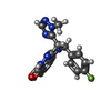  Mass: 380.351 Da / Num. of mol.: 2 / Source method: obtained synthetically / Formula: C19H14F2N6O Mass: 380.351 Da / Num. of mol.: 2 / Source method: obtained synthetically / Formula: C19H14F2N6O#3: Chemical |   Mass: 92.094 Da / Num. of mol.: 3 / Source method: obtained synthetically / Formula: C3H8O3 Mass: 92.094 Da / Num. of mol.: 3 / Source method: obtained synthetically / Formula: C3H8O3#4: Water | ChemComp-HOH / |  Mass: 18.015 Da / Num. of mol.: 143 / Source method: isolated from a natural source / Formula: H2O Mass: 18.015 Da / Num. of mol.: 143 / Source method: isolated from a natural source / Formula: H2OSequence details | PRO to HIS conflict in unp entry Q9UGN5 | |
|---|
-Experimental details
-Experiment
| Experiment | Method: X-RAY DIFFRACTION / Number of used crystals: 1 |
|---|
- Sample preparation
| Crystal | Density Matthews: 2.21 Å3/Da / Density % sol: 44.4 % |
|---|---|
| Crystal grow | Temperature: 289 K / Method: vapor diffusion / pH: 7.5 Details: (W/V) POLYETHYLENE GLYCOL 3350, 333 mM SODIUM CHLORIDE. PH range: 7.5 |
-Data collection
| Diffraction | Mean temperature: 100 K |
|---|---|
| Diffraction source | Source: SYNCHROTRON / Site: SSRL  / Beamline: BL7-1 / Wavelength: 1.097 Å / Beamline: BL7-1 / Wavelength: 1.097 Å |
| Detector | Type: ADSC QUANTUM 315 / Detector: CCD / Date: Jul 19, 2011 |
| Radiation | Protocol: SINGLE WAVELENGTH / Monochromatic (M) / Laue (L): M / Scattering type: x-ray |
| Radiation wavelength | Wavelength: 1.097 Å / Relative weight: 1 |
| Reflection | Resolution: 2.5→67.33 Å / Num. obs: 22773 / % possible obs: 91.9 % / Observed criterion σ(I): -3 / Redundancy: 2 % / Biso Wilson estimate: 25.67 Å2 / Rmerge(I) obs: 0.123 / Net I/σ(I): 7.02 |
| Reflection shell | Resolution: 2.5→2.56 Å / Rmerge(I) obs: 0.457 / Mean I/σ(I) obs: 1.8 / % possible all: 91.3 |
- Processing
| Software |
| ||||||||||||||||||||||||||||||||||||||||||||||||||||||||||||||||||||||||||||||||||||||||||||||||||||||||||||||||||||||||||||||||||||||||||||||||||||||||||||||||||||||||||||||||||||||
|---|---|---|---|---|---|---|---|---|---|---|---|---|---|---|---|---|---|---|---|---|---|---|---|---|---|---|---|---|---|---|---|---|---|---|---|---|---|---|---|---|---|---|---|---|---|---|---|---|---|---|---|---|---|---|---|---|---|---|---|---|---|---|---|---|---|---|---|---|---|---|---|---|---|---|---|---|---|---|---|---|---|---|---|---|---|---|---|---|---|---|---|---|---|---|---|---|---|---|---|---|---|---|---|---|---|---|---|---|---|---|---|---|---|---|---|---|---|---|---|---|---|---|---|---|---|---|---|---|---|---|---|---|---|---|---|---|---|---|---|---|---|---|---|---|---|---|---|---|---|---|---|---|---|---|---|---|---|---|---|---|---|---|---|---|---|---|---|---|---|---|---|---|---|---|---|---|---|---|---|---|---|---|---|
| Refinement | Method to determine structure: MOLECULAR REPLACEMENT Starting model: 3KCZ Resolution: 2.5→67.33 Å / Cor.coef. Fo:Fc: 0.91 / Cor.coef. Fo:Fc free: 0.848 / SU B: 22.703 / SU ML: 0.258 / Cross valid method: THROUGHOUT / σ(F): 0 / ESU R: 2.366 / ESU R Free: 0.372 / Stereochemistry target values: MAXIMUM LIKELIHOOD Details: U VALUES : WITH TLS ADDED HYDROGENS HAVE BEEN ADDED IN THE RIDING POSITIONS
| ||||||||||||||||||||||||||||||||||||||||||||||||||||||||||||||||||||||||||||||||||||||||||||||||||||||||||||||||||||||||||||||||||||||||||||||||||||||||||||||||||||||||||||||||||||||
| Solvent computation | Ion probe radii: 0.8 Å / Shrinkage radii: 0.8 Å / VDW probe radii: 1.2 Å / Solvent model: MASK | ||||||||||||||||||||||||||||||||||||||||||||||||||||||||||||||||||||||||||||||||||||||||||||||||||||||||||||||||||||||||||||||||||||||||||||||||||||||||||||||||||||||||||||||||||||||
| Displacement parameters | Biso mean: 20.88 Å2
| ||||||||||||||||||||||||||||||||||||||||||||||||||||||||||||||||||||||||||||||||||||||||||||||||||||||||||||||||||||||||||||||||||||||||||||||||||||||||||||||||||||||||||||||||||||||
| Refinement step | Cycle: 1 / Resolution: 2.5→67.33 Å
| ||||||||||||||||||||||||||||||||||||||||||||||||||||||||||||||||||||||||||||||||||||||||||||||||||||||||||||||||||||||||||||||||||||||||||||||||||||||||||||||||||||||||||||||||||||||
| Refine LS restraints |
|