[English] 日本語
Yorodumi
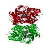
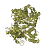
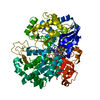
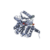
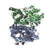
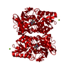
- PDB-5d84: Staphyloferrin B precursor biosynthetic enzyme SbnA bound to PLP -

+
Open data


ID or keywords:

Loading...

-
Basic information

Entry
Database: PDB / ID: 5d84
TitleStaphyloferrin B precursor biosynthetic enzyme SbnA bound to PLP
ComponentsProbable siderophore biosynthesis protein SbnA
KeywordsBIOSYNTHETIC PROTEIN / siderophore / iron / plp
Function / homology
Function and homology information


N-(2-amino-2-carboxyethyl)-L-glutamate synthase / cysteine biosynthetic process from serine / transferase activity, transferring alkyl or aryl (other than methyl) groups
Similarity search - Function
2,3-diaminopropionate biosynthesis protein SbnA / Cysteine synthase/cystathionine beta-synthase, pyridoxal-phosphate attachment site / Cysteine synthase/cystathionine beta-synthase P-phosphate attachment site. / Rossmann fold - #1100 / Pyridoxal-phosphate dependent enzyme / Tryptophan synthase beta subunit-like PLP-dependent enzyme / Pyridoxal-phosphate dependent enzyme / Rossmann fold / 3-Layer(aba) Sandwich / Alpha Beta
Similarity search - Domain/homology
PYRIDOXAL-5'-PHOSPHATE / N-(2-amino-2-carboxyethyl)-L-glutamate synthase
Similarity search - Component
Biological speciesStaphylococcus aureus (bacteria)
MethodX-RAY DIFFRACTION / SYNCHROTRON / MOLECULAR REPLACEMENT / molecular replacement / Resolution: 1.45 Å
AuthorsGrigg, J.C. / Kobylarz, M.J. / Liu, Y. / Lee, M.S.F. / Heinrichs, D.E. / Murphy, M.E.P.
Funding support Canada, 1items
OrganizationGrant numberCountry
Canadian Institutes of Health Research (CIHR)MOP-102596 Canada
CitationJournal: Biochemistry / Year: 2016
Title: Deciphering the Substrate Specificity of SbnA, the Enzyme Catalyzing the First Step in Staphyloferrin B Biosynthesis.
Authors: Kobylarz, M.J. / Grigg, J.C. / Liu, Y. / Lee, M.S. / Heinrichs, D.E. / Murphy, M.E.
History
DepositionAug 15, 2015Deposition site: RCSB / Processing site: RCSB
Revision 1.0Feb 3, 2016Provider: repository / Type: Initial release
Revision 1.1Feb 24, 2016Group: Database references
Revision 1.2Jan 8, 2020Group: Author supporting evidence / Database references / Derived calculations
Category: citation / pdbx_audit_support / pdbx_struct_oper_list
Item: _citation.journal_id_CSD / _pdbx_audit_support.funding_organization / _pdbx_struct_oper_list.symmetry_operation

-
Structure visualization

Structure viewerMolecule:
MolmilJmol/JSmol

Downloads & links

-
Assembly

Deposited unit
A: Probable siderophore biosynthesis protein SbnA
hetero molecules


Theoretical massNumber of molelcules
Total (without water)36,2123
Polymers35,9401
Non-polymers2712
Water6,557364
1
A: Probable siderophore biosynthesis protein SbnA
hetero molecules

A: Probable siderophore biosynthesis protein SbnA
hetero molecules


Theoretical massNumber of molelcules
Total (without water)72,4236
Polymers71,8802
Non-polymers5434
Water362
TypeNameSymmetry operationNumber
identity operation1_555x,y,z1
crystal symmetry operation2_655-x+1,-y,z1
Buried area4350 Å2
ΔGint-26 kcal/mol
Surface area25070 Å2
MethodPISA
Unit cell
Length a, b, c (Å)56.080, 115.828, 45.138
Angle α, β, γ (deg.)90.000, 90.000, 90.000
Int Tables number18
Space group name H-MP21212
Components on special symmetry positions
IDModelComponents
11A-1236-

HOH

-
Components

#1: Protein Probable siderophore biosynthesis protein SbnA


Mass: 35940.164 Da / Num. of mol.: 1
Source method: isolated from a genetically manipulated source
Source: (gene. exp.) Staphylococcus aureus (bacteria) / Strain: Newman / Gene: sbnA, NWMN_0060 / Plasmid: pET28a / Production host: Escherichia coli (E. coli) / Strain (production host): BL21(DE3) / References: UniProt: A6QDA0
#2: Chemical ChemComp-PLP / PYRIDOXAL-5'-PHOSPHATE / VITAMIN B6 Phosphate / Pyridoxal phosphate


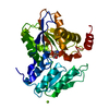
Mass: 247.142 Da / Num. of mol.: 1 / Source method: obtained synthetically / Formula: C8H10NO6P
#3: Chemical ChemComp-MG / MAGNESIUM ION


Mass: 24.305 Da / Num. of mol.: 1 / Source method: obtained synthetically / Formula: Mg
#4: Water ChemComp-HOH / water / Water


Mass: 18.015 Da / Num. of mol.: 364 / Source method: isolated from a natural source / Formula: H2O

-
Experimental details

-
Experiment

ExperimentMethod: X-RAY DIFFRACTION / Number of used crystals: 1

-
Sample preparation

CrystalDensity Matthews: 2.04 Å3/Da / Density % sol: 39.69 %
Crystal growTemperature: 293 K / Method: vapor diffusion, hanging drop / pH: 8.5 / Details: 0.1 M Tris, pH 8.5, 0.2 M MgCl2, 20-25% PEG 3350

-
Data collection

DiffractionMean temperature: 100 K
Diffraction sourceSource: SYNCHROTRON / Site: CLSI / Beamline: 08ID-1 / Wavelength: 0.97952 Å
DetectorType: RAYONIX MX300HE / Detector: CCD / Date: Dec 15, 2010
Details: Vertical Focusing Mirror: ultra-low expansion (ULE) titanium siliicate flat mirror with Pt, Uncoated, and Pd strips
RadiationMonochromator: ACCEL/BRUKER double crystal monochromator (DCM), featuring indirectly cryo-cooled first crystal and sagittally focusing second crystal
Protocol: SINGLE WAVELENGTH / Monochromatic (M) / Laue (L): M / Scattering type: x-ray
Radiation wavelengthWavelength: 0.97952 Å / Relative weight: 1
ReflectionResolution: 1.45→50 Å / Num. obs: 51631 / % possible obs: 97.3 % / Redundancy: 5.9 % / Rmerge(I) obs: 0.055 / Χ2: 1.006 / Net I/av σ(I): 28.097 / Net I/σ(I): 13 / Num. measured all: 302661
Reflection shell

Diffraction-ID: 1 / Rejects: 0

Resolution (Å)Redundancy (%)Rmerge(I) obsNum. unique allΧ2% possible all
1.45-1.55.60.41349541.01994.7
1.5-1.565.70.31849420.99994.6
1.56-1.635.70.25949571.00394.8
1.63-1.725.70.19850051.01795.6
1.72-1.835.70.15450990.95997.3
1.83-1.975.80.10952280.93698.7
1.97-2.1760.0752690.99399.7
2.17-2.486.20.05553171.00499.8
2.48-3.126.20.04153770.991100
3.12-5060.02854831.13197.5

-
Phasing

PhasingMethod: molecular replacement
Phasing MR
Highest resolutionLowest resolution
Rotation1.8 Å40.12 Å
Translation1.8 Å40.12 Å

-
Processing

Software
NameVersionClassification
HKL-2000data scaling
PHENIX2.2.4phasing
REFMAC5.8.0073refinement
PDB_EXTRACT3.15data extraction
HKL-2000data reduction
RefinementMethod to determine structure: MOLECULAR REPLACEMENT / Resolution: 1.45→50 Å / Cor.coef. Fo:Fc: 0.98 / Cor.coef. Fo:Fc free: 0.965 / SU B: 2.585 / SU ML: 0.044 / Cross valid method: THROUGHOUT / σ(F): 0 / ESU R: 0.069 / ESU R Free: 0.065 / Stereochemistry target values: MAXIMUM LIKELIHOOD / Details: HYDROGENS HAVE BEEN USED IF PRESENT IN THE INPUT
RfactorNum. reflection% reflectionSelection details
Rfree0.1753 2598 5.1 %RANDOM
Rwork0.1239 ---
obs0.1265 48724 96.82 %-
Solvent computationIon probe radii: 0.8 Å / Shrinkage radii: 0.8 Å / VDW probe radii: 1.2 Å / Solvent model: MASK
Displacement parametersBiso max: 104.86 Å2 / Biso mean: 18.813 Å2 / Biso min: 7.92 Å2
Baniso -1Baniso -2Baniso -3
1--0.25 Å20 Å20 Å2
2--0.59 Å20 Å2
3----0.35 Å2
Refinement stepCycle: final / Resolution: 1.45→50 Å
ProteinNucleic acidLigandSolventTotal
Num. atoms2456 0 16 364 2836
Biso mean--12.9 32.78 -
Num. residues----318
Refine LS restraints
Refine-IDTypeDev idealDev ideal targetNumber
X-RAY DIFFRACTIONr_bond_refined_d0.0120.0192714
X-RAY DIFFRACTIONr_angle_refined_deg1.5151.9693705
X-RAY DIFFRACTIONr_dihedral_angle_1_deg5.4365358
X-RAY DIFFRACTIONr_dihedral_angle_2_deg36.92325.089112
X-RAY DIFFRACTIONr_dihedral_angle_3_deg12.46215492
X-RAY DIFFRACTIONr_dihedral_angle_4_deg15.1781512
X-RAY DIFFRACTIONr_chiral_restr0.0920.2416
X-RAY DIFFRACTIONr_gen_planes_refined0.0090.0212061
X-RAY DIFFRACTIONr_mcbond_it2.6271.4851375
X-RAY DIFFRACTIONr_mcangle_it3.3272.2451752
X-RAY DIFFRACTIONr_scbond_it3.3541.7821339
X-RAY DIFFRACTIONr_rigid_bond_restr3.04932714
X-RAY DIFFRACTIONr_sphericity_free42.449589
X-RAY DIFFRACTIONr_sphericity_bonded16.27852922
LS refinement shellResolution: 1.45→1.488 Å / Total num. of bins used: 20
RfactorNum. reflection% reflection
Rfree0.259 192 -
Rwork0.19 3421 -
all-3613 -
obs--94.53 %

+
About Yorodumi

-
News

-
Feb 9, 2022. New format data for meta-information of EMDB entries

New format data for meta-information of EMDB entries

  • Version 3 of the EMDB header file is now the official format.
  • The previous official version 1.9 will be removed from the archive.

Related info.:EMDB header

External links:wwPDB to switch to version 3 of the EMDB data model

-
Aug 12, 2020. Covid-19 info

Covid-19 info

URL: https://pdbj.org/emnavi/covid19.php

New page: Covid-19 featured information page in EM Navigator.

Related info.:Covid-19 info / Mar 5, 2020. Novel coronavirus structure data

+
Mar 5, 2020. Novel coronavirus structure data

Novel coronavirus structure data

Related info.:Yorodumi Speices / Aug 12, 2020. Covid-19 info

External links:COVID-19 featured content - PDBj / Molecule of the Month (242):Coronavirus Proteases

+
Jan 31, 2019. EMDB accession codes are about to change! (news from PDBe EMDB page)

EMDB accession codes are about to change! (news from PDBe EMDB page)

  • The allocation of 4 digits for EMDB accession codes will soon come to an end. Whilst these codes will remain in use, new EMDB accession codes will include an additional digit and will expand incrementally as the available range of codes is exhausted. The current 4-digit format prefixed with “EMD-” (i.e. EMD-XXXX) will advance to a 5-digit format (i.e. EMD-XXXXX), and so on. It is currently estimated that the 4-digit codes will be depleted around Spring 2019, at which point the 5-digit format will come into force.
  • The EM Navigator/Yorodumi systems omit the EMD- prefix.

Related info.:Q: What is EMD? / ID/Accession-code notation in Yorodumi/EM Navigator

External links:EMDB Accession Codes are Changing Soon! / Contact to PDBj

+
Jul 12, 2017. Major update of PDB

Major update of PDB

  • wwPDB released updated PDB data conforming to the new PDBx/mmCIF dictionary.
  • This is a major update changing the version number from 4 to 5, and with Remediation, in which all the entries are updated.
  • In this update, many items about electron microscopy experimental information are reorganized (e.g. em_software).
  • Now, EM Navigator and Yorodumi are based on the updated data.

External links:wwPDB Remediation / Enriched Model Files Conforming to OneDep Data Standards Now Available in the PDB FTP Archive

-
Yorodumi

Thousand views of thousand structures

  • Yorodumi is a browser for structure data from EMDB, PDB, SASBDB, etc.
  • This page is also the successor to EM Navigator detail page, and also detail information page/front-end page for Omokage search.
  • The word "yorodu" (or yorozu) is an old Japanese word meaning "ten thousand". "mi" (miru) is to see.

Related info.:EMDB / PDB / SASBDB / Comparison of 3 databanks / Yorodumi Search / Aug 31, 2016. New EM Navigator & Yorodumi / Yorodumi Papers / Jmol/JSmol / Function and homology information / Changes in new EM Navigator and Yorodumi

Read more