 Movie
Movie Controller

[English] 日本語
 Yorodumi
- PDB-1olz: The ligand-binding face of the semaphorins revealed by the high r... -
+ Open data

- Basic information
| Entry | Database: PDB / ID: 1olz | ||||||
|---|---|---|---|---|---|---|---|
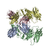
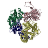
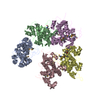
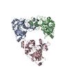
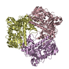
| Title | The ligand-binding face of the semaphorins revealed by the high resolution crystal structure of SEMA4D | ||||||
 Components Components | SEMAPHORIN 4D | ||||||
 Keywords Keywords | DEVELOPMENTAL PROTEIN / CD100 / SEMAPHORIN / BETA-PROPELLER / PSI DOMAIN / IG-LIKE DOMAIN / EXTRACELLULAR RECEPTOR / NEUROGENESIS / GLYCOPROTEIN DEVELOPMENTAL PROTEIN / STRUCTURAL PROTEOMICS IN EUROPE / SPINE / STRUCTURAL GENOMICS | ||||||
| Function / homology |  Function and homology information Function and homology informationleukocyte aggregation / positive regulation of inhibitory synapse assembly / semaphorin receptor binding / regulation of cell projection organization / Other semaphorin interactions / bone trabecula morphogenesis / positive regulation of collateral sprouting / semaphorin receptor complex / neuropilin binding / chemorepellent activity ...leukocyte aggregation / positive regulation of inhibitory synapse assembly / semaphorin receptor binding / regulation of cell projection organization / Other semaphorin interactions / bone trabecula morphogenesis / positive regulation of collateral sprouting / semaphorin receptor complex / neuropilin binding / chemorepellent activity / negative regulation of cell adhesion / Sema4D induced cell migration and growth-cone collapse / Sema4D mediated inhibition of cell attachment and migration / ossification involved in bone maturation / neural crest cell migration / negative chemotaxis / regulation of dendrite morphogenesis / positive regulation of Rho protein signal transduction / ciliary transition zone / semaphorin-plexin signaling pathway / positive regulation of GTPase activity / negative regulation of osteoblast differentiation / axon guidance / positive regulation of protein phosphorylation / GABA-ergic synapse / transmembrane signaling receptor activity / regulation of cell shape / signaling receptor activity / postsynaptic membrane / positive regulation of phosphatidylinositol 3-kinase/protein kinase B signal transduction / cell adhesion / immune response / positive regulation of cell migration / ciliary basal body / receptor ligand activity / signaling receptor binding / centrosome / negative regulation of apoptotic process / negative regulation of transcription by RNA polymerase II / : / nucleoplasm / identical protein binding / plasma membrane Similarity search - Function | ||||||
| Biological species |  Homo sapiens (human) Homo sapiens (human) | ||||||
| Method |  X-RAY DIFFRACTION / SYNCHROTRON / MAD / Resolution: 2 Å X-RAY DIFFRACTION / SYNCHROTRON / MAD / Resolution: 2 Å | ||||||
 Authors Authors | Love, C.A. / Harlos, K. / Mavaddat, N. / Davis, S.J. / Stuart, D.I. / Jones, E.Y. / Esnouf, R.M. | ||||||
 Citation Citation | Journal: Nat.Struct.Biol. / Year: 2003 Title: The Ligand-Binding Face of the Semaphorins Revealed by the High-Resolution Crystal Structure of Sema4D Authors: Love, C.A. / Harlos, K. / Mavaddat, N. / Davis, S.J. / Stuart, D.I. / Jones, E.Y. / Esnouf, R.M. | ||||||
| History |
| ||||||
| Remark 700 | SHEET THE SHEET STRUCTURE OF THIS MOLECULE IS BIFURCATED. IN ORDER TO REPRESENT THIS FEATURE IN ... SHEET THE SHEET STRUCTURE OF THIS MOLECULE IS BIFURCATED. IN ORDER TO REPRESENT THIS FEATURE IN THE SHEET RECORDS BELOW, TWO SHEETS ARE DEFINED. |
- Structure visualization
| Structure viewer | Molecule: MolmilJmol/JSmol |
|---|
- Downloads & links
-Download
| PDBx/mmCIF format | 1olz.cif.gz | 272.2 KB | Display | PDBx/mmCIF format |
|---|---|---|---|---|
| PDB format | pdb1olz.ent.gz | 217.9 KB | Display | PDB format |
| PDBx/mmJSON format | 1olz.json.gz | Tree view | PDBx/mmJSON format | |
| Others |  Other downloads Other downloads |
-Validation report
| Arichive directory | https://data.pdbj.org/pub/pdb/validation_reports/ol/1olzftp://data.pdbj.org/pub/pdb/validation_reports/ol/1olz | HTTPS FTP |
|---|
-Related structure data
| Similar structure data |
|---|


-Links
 PDBj
PDBj


- Assembly
| Deposited unit | 
| ||||||||
|---|---|---|---|---|---|---|---|---|---|
| 1 |
| ||||||||
| Unit cell |
| ||||||||
| Noncrystallographic symmetry (NCS) | NCS oper: (Code: given Matrix: (0.50294, -0.85108, -0.15071), Vector: |
-Components
| #1: Antibody | Mass: 74315.547 Da / Num. of mol.: 2 / Fragment: SOLUBLE EXTRACELLULAR FRAGMENT, RESIDUES 22-677 Source method: isolated from a genetically manipulated source Source: (gene. exp.) Homo sapiens (human) / Cell line (production host): CHO / Production host:   Cricetulus griseus (Chinese hamster) / Variant (production host): LEC3.2.8.1 / References: UniProt: Q92854 Cricetulus griseus (Chinese hamster) / Variant (production host): LEC3.2.8.1 / References: UniProt: Q92854#2: Water | ChemComp-HOH / |  Mass: 18.015 Da / Num. of mol.: 841 / Source method: isolated from a natural source / Formula: H2O Mass: 18.015 Da / Num. of mol.: 841 / Source method: isolated from a natural source / Formula: H2OHas protein modification | Y | |
|---|
-Experimental details
-Experiment
| Experiment | Method: X-RAY DIFFRACTION / Number of used crystals: 1 |
|---|
- Sample preparation
| Crystal | Density Matthews: 2.89 Å3/Da / Density % sol: 57 % | ||||||||||||||||||||||||||||
|---|---|---|---|---|---|---|---|---|---|---|---|---|---|---|---|---|---|---|---|---|---|---|---|---|---|---|---|---|---|
| Crystal grow | pH: 7.5 / Details: 0.2 M AMMONIUM FLORIDE, 20% PEG 3350, pH 7.50 | ||||||||||||||||||||||||||||
| Crystal grow | *PLUS pH: 8 / Method: vapor diffusion, sitting drop | ||||||||||||||||||||||||||||
| Components of the solutions | *PLUS
|
-Data collection
| Diffraction | Mean temperature: 100 K |
|---|---|
| Diffraction source | Source: SYNCHROTRON / Site: ESRF  / Beamline: ID29 / Wavelength: 0.975 / Beamline: ID29 / Wavelength: 0.975 |
| Detector | Type: ADSC CCD / Detector: CCD / Date: Jun 20, 2003 / Details: TOROIDAL MIRRORS |
| Radiation | Monochromator: SI(111) / Protocol: SINGLE WAVELENGTH / Monochromatic (M) / Laue (L): M / Scattering type: x-ray |
| Radiation wavelength | Wavelength: 0.975 Å / Relative weight: 1 |
| Reflection | Resolution: 2→20 Å / Num. obs: 108424 / % possible obs: 97.2 % / Redundancy: 3.5 % / Biso Wilson estimate: 28.5 Å2 / Rmerge(I) obs: 0.099 / Net I/σ(I): 17.6 |
| Reflection shell | Resolution: 2→2.07 Å / Redundancy: 3 % / Rmerge(I) obs: 0.801 / Mean I/σ(I) obs: 1.8 / % possible all: 90.9 |
| Reflection | *PLUS Highest resolution: 2 Å / Num. measured all: 375205 / Rmerge(I) obs: 0.099 |
| Reflection shell | *PLUS % possible obs: 90.9 % / Mean I/σ(I) obs: 1.8 |
- Processing
| Software |
| ||||||||||||||||||||||||||||||||||||||||||||||||||||||||||||||||||||||||||||||||
|---|---|---|---|---|---|---|---|---|---|---|---|---|---|---|---|---|---|---|---|---|---|---|---|---|---|---|---|---|---|---|---|---|---|---|---|---|---|---|---|---|---|---|---|---|---|---|---|---|---|---|---|---|---|---|---|---|---|---|---|---|---|---|---|---|---|---|---|---|---|---|---|---|---|---|---|---|---|---|---|---|---|
| Refinement | Method to determine structure: MAD / Resolution: 2→20 Å / Data cutoff high absF: 1000000 / Data cutoff low absF: 0.001 / Isotropic thermal model: RESTAINED / Cross valid method: THROUGHOUT / σ(F): 0 Details: THE PROTEIN WAS PRODUCED IN A EUKARYOTIC EXPRESSION SYSTEM WHICH IS THEREFORE NOT FULLY SELENO-METHIONINE LABELLED SINCE THE EXACT COMPOSITION IS UNKNOWN, WE HAVE USED A CONSERVATIVE ...Details: THE PROTEIN WAS PRODUCED IN A EUKARYOTIC EXPRESSION SYSTEM WHICH IS THEREFORE NOT FULLY SELENO-METHIONINE LABELLED SINCE THE EXACT COMPOSITION IS UNKNOWN, WE HAVE USED A CONSERVATIVE APPROACH IN REFINEMENT AND TREATED THE RESIDUES AS STANDARD METHIONINE RESIDUES ALLOWING B FACTORS TO COMPENSATE FOR OCCUPANCY.
| ||||||||||||||||||||||||||||||||||||||||||||||||||||||||||||||||||||||||||||||||
| Refinement step | Cycle: LAST / Resolution: 2→20 Å
| ||||||||||||||||||||||||||||||||||||||||||||||||||||||||||||||||||||||||||||||||
| Refine LS restraints |
| ||||||||||||||||||||||||||||||||||||||||||||||||||||||||||||||||||||||||||||||||
| LS refinement shell | Resolution: 2→2.09 Å / Total num. of bins used: 8
| ||||||||||||||||||||||||||||||||||||||||||||||||||||||||||||||||||||||||||||||||
| Xplor file |
| ||||||||||||||||||||||||||||||||||||||||||||||||||||||||||||||||||||||||||||||||
| Refinement | *PLUS Highest resolution: 2 Å / Lowest resolution: 20 Å / % reflection Rfree: 5 % | ||||||||||||||||||||||||||||||||||||||||||||||||||||||||||||||||||||||||||||||||
| Solvent computation | *PLUS | ||||||||||||||||||||||||||||||||||||||||||||||||||||||||||||||||||||||||||||||||
| Displacement parameters | *PLUS | ||||||||||||||||||||||||||||||||||||||||||||||||||||||||||||||||||||||||||||||||
| Refine LS restraints | *PLUS
|
