 Movie
Movie Controller


+ Open data

- Basic information
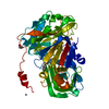
| Entry | Database: PDB / ID: 6fyk | ||||||
|---|---|---|---|---|---|---|---|
| Title | X-Ray structure of CLK2-KD(136-496)/Indazole1 at 2.39A | ||||||
 Components Components | Dual specificity protein kinase CLK2 | ||||||
 Keywords Keywords | TRANSFERASE / SPLICING / PHOSPHOTRANSFERASE | ||||||
| Function / homology |  Function and homology information Function and homology informationdual-specificity kinase / response to ionizing radiation / regulation of RNA splicing / negative regulation of gluconeogenesis / protein serine/threonine/tyrosine kinase activity / protein autophosphorylation / protein tyrosine kinase activity / protein phosphorylation / nuclear speck / nuclear body ...dual-specificity kinase / response to ionizing radiation / regulation of RNA splicing / negative regulation of gluconeogenesis / protein serine/threonine/tyrosine kinase activity / protein autophosphorylation / protein tyrosine kinase activity / protein phosphorylation / nuclear speck / nuclear body / protein serine kinase activity / protein serine/threonine kinase activity / nucleoplasm / ATP binding / identical protein binding / nucleus Similarity search - Function | ||||||
| Biological species |  Homo sapiens (human) Homo sapiens (human) | ||||||
| Method |  X-RAY DIFFRACTION / SYNCHROTRON / MOLECULAR REPLACEMENT / Resolution: 2.39 Å X-RAY DIFFRACTION / SYNCHROTRON / MOLECULAR REPLACEMENT / Resolution: 2.39 Å | ||||||
 Authors Authors | Kallen, J. | ||||||
 Citation Citation | Journal: ChemMedChem / Year: 2018 Title: X-ray Structures and Feasibility Assessment of CLK2 Inhibitors for Phelan-McDermid Syndrome. Authors: Kallen, J. / Bergsdorf, C. / Arnaud, B. / Bernhard, M. / Brichet, M. / Cobos-Correa, A. / Elhajouji, A. / Freuler, F. / Galimberti, I. / Guibourdenche, C. / Haenni, S. / Holzinger, S. / ...Authors: Kallen, J. / Bergsdorf, C. / Arnaud, B. / Bernhard, M. / Brichet, M. / Cobos-Correa, A. / Elhajouji, A. / Freuler, F. / Galimberti, I. / Guibourdenche, C. / Haenni, S. / Holzinger, S. / Hunziker, J. / Izaac, A. / Kaufmann, M. / Leder, L. / Martus, H.J. / von Matt, P. / Polyakov, V. / Roethlisberger, P. / Roma, G. / Stiefl, N. / Uteng, M. / Lerchner, A. | ||||||
| History |
|
- Structure visualization
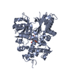
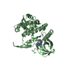
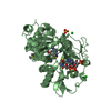
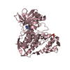
| Structure viewer | Molecule: MolmilJmol/JSmol |
|---|
- Downloads & links
-Download
| PDBx/mmCIF format | 6fyk.cif.gz | 234.5 KB | Display | PDBx/mmCIF format |
|---|---|---|---|---|
| PDB format | pdb6fyk.ent.gz | 187.4 KB | Display | PDB format |
| PDBx/mmJSON format | 6fyk.json.gz | Tree view | PDBx/mmJSON format | |
| Others |  Other downloads Other downloads |
-Validation report
| Arichive directory | https://data.pdbj.org/pub/pdb/validation_reports/fy/6fykftp://data.pdbj.org/pub/pdb/validation_reports/fy/6fyk | HTTPS FTP |
|---|
-Related structure data
| Related structure data |  6fyiSC  6fylC  6fyoC  6fypC  6fyrC  6fyvC S: Starting model for refinement C: citing same article ( |
|---|---|
| Similar structure data |


-Links
 PDBj
PDBj- Assembly
| Deposited unit | 
| ||||||||
|---|---|---|---|---|---|---|---|---|---|
| 1 | 
| ||||||||
| 2 | 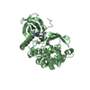
| ||||||||
| 3 | 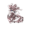
| ||||||||
| Unit cell |
| ||||||||
| Components on special symmetry positions |
|
-Components
| #1: Protein | Mass: 42767.500 Da / Num. of mol.: 3 / Fragment: kinase domain Source method: isolated from a genetically manipulated source Source: (gene. exp.) Homo sapiens (human) / Gene: CLK2 / Production host:  #2: Chemical | 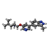  Mass: 324.420 Da / Num. of mol.: 3 / Source method: obtained synthetically / Formula: C19H24N4O Mass: 324.420 Da / Num. of mol.: 3 / Source method: obtained synthetically / Formula: C19H24N4O#3: Water | ChemComp-HOH / |  Mass: 18.015 Da / Num. of mol.: 466 / Source method: isolated from a natural source / Formula: H2O Mass: 18.015 Da / Num. of mol.: 466 / Source method: isolated from a natural source / Formula: H2O |
|---|
-Experimental details
-Experiment
| Experiment | Method: X-RAY DIFFRACTION / Number of used crystals: 1 |
|---|
- Sample preparation
| Crystal | Density Matthews: 2.38 Å3/Da / Density % sol: 48.29 % |
|---|---|
| Crystal grow | Temperature: 298 K / Method: vapor diffusion, sitting drop / pH: 8.5 Details: 20% PEG2000, 0.2M trimethylamine n-oxide, 0.1M TRIS |
-Data collection
| Diffraction | Mean temperature: 100 K |
|---|---|
| Diffraction source | Source: SYNCHROTRON / Site: SLS  / Beamline: X10SA / Wavelength: 0.99986 Å / Beamline: X10SA / Wavelength: 0.99986 Å |
| Detector | Type: DECTRIS PILATUS 6M / Detector: PIXEL / Date: Oct 1, 2016 |
| Radiation | Monochromator: SI 111 channel / Protocol: SINGLE WAVELENGTH / Monochromatic (M) / Laue (L): M / Scattering type: x-ray |
| Radiation wavelength | Wavelength: 0.99986 Å / Relative weight: 1 |
| Reflection | Resolution: 2.39→19.68 Å / Num. obs: 47518 / % possible obs: 99.8 % / Redundancy: 10 % / Biso Wilson estimate: 37.4 Å2 / CC1/2: 0.998 / Rmerge(I) obs: 0.163 / Rrim(I) all: 0.171 / Net I/σ(I): 14.4 |
| Reflection shell | Resolution: 2.39→2.45 Å / Redundancy: 9.5 % / Rmerge(I) obs: 0.927 / Mean I/σ(I) obs: 2.9 / CC1/2: 0.859 / Rrim(I) all: 0.981 / % possible all: 100 |
- Processing
| Software |
| |||||||||||||||||||||||||||||||||||||||||||||
|---|---|---|---|---|---|---|---|---|---|---|---|---|---|---|---|---|---|---|---|---|---|---|---|---|---|---|---|---|---|---|---|---|---|---|---|---|---|---|---|---|---|---|---|---|---|---|
| Refinement | Method to determine structure: MOLECULAR REPLACEMENT Starting model: 6FYI Resolution: 2.39→19.68 Å / Cor.coef. Fo:Fc: 0.945 / Cor.coef. Fo:Fc free: 0.923 / SU B: 7.441 / SU ML: 0.171 / SU R Cruickshank DPI: 0.5139 / Cross valid method: THROUGHOUT / σ(F): 0 / ESU R: 0.514 / ESU R Free: 0.252 Details: HYDROGENS HAVE BEEN USED IF PRESENT IN THE INPUT U VALUES : REFINED INDIVIDUALLY
| |||||||||||||||||||||||||||||||||||||||||||||
| Solvent computation | Ion probe radii: 0.8 Å / Shrinkage radii: 0.8 Å / VDW probe radii: 1.2 Å | |||||||||||||||||||||||||||||||||||||||||||||
| Displacement parameters | Biso max: 155.99 Å2 / Biso mean: 36.307 Å2 / Biso min: 14.79 Å2
| |||||||||||||||||||||||||||||||||||||||||||||
| Refinement step | Cycle: final / Resolution: 2.39→19.68 Å
| |||||||||||||||||||||||||||||||||||||||||||||
| Refine LS restraints |
| |||||||||||||||||||||||||||||||||||||||||||||
| LS refinement shell | Resolution: 2.39→2.451 Å / Rfactor Rfree error: 0 / Total num. of bins used: 20
|
