 Movie
Movie Controller


+ Open data

- Basic information
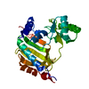
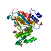
| Entry | Database: PDB / ID: 1ojq | ||||||
|---|---|---|---|---|---|---|---|
| Title | The crystal structure of C3stau2 from S. aureus | ||||||
 Components Components | ADP-RIBOSYLTRANSFERASE | ||||||
 Keywords Keywords | TRANSFERASE / ADP-RIBOSYLTRANSFERASE | ||||||
| Function / homology |  Function and homology information Function and homology informationNAD+-protein mono-ADP-ribosyltransferase activity / nucleotide binding / extracellular region Similarity search - Function | ||||||
| Biological species |   STAPHYLOCOCCUS AUREUS (bacteria) STAPHYLOCOCCUS AUREUS (bacteria) | ||||||
| Method |  X-RAY DIFFRACTION / SYNCHROTRON / MOLECULAR REPLACEMENT / Resolution: 1.68 Å X-RAY DIFFRACTION / SYNCHROTRON / MOLECULAR REPLACEMENT / Resolution: 1.68 Å | ||||||
 Authors Authors | Evans, H.R. / Sutton, J.M. / Holloway, D.E. / Ayriss, J. / Shone, C.C. / Acharya, K.R. | ||||||
 Citation Citation | Journal: J.Biol.Chem. / Year: 2003 Title: The Crystal Structure of C3Stau2 from Staphylococcus Aureus and its Complex with Nad Authors: Evans, H.R. / Sutton, J.M. / Holloway, D.E. / Ayriss, J. / Shone, C.C. / Acharya, K.R. | ||||||
| History |
|
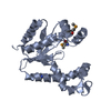
- Structure visualization
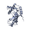
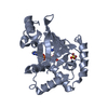
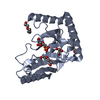
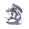
| Structure viewer | Molecule: MolmilJmol/JSmol |
|---|
- Downloads & links
-Download
| PDBx/mmCIF format | 1ojq.cif.gz | 59.3 KB | Display | PDBx/mmCIF format |
|---|---|---|---|---|
| PDB format | pdb1ojq.ent.gz | 42.8 KB | Display | PDB format |
| PDBx/mmJSON format | 1ojq.json.gz | Tree view | PDBx/mmJSON format | |
| Others |  Other downloads Other downloads |
-Validation report
| Arichive directory | https://data.pdbj.org/pub/pdb/validation_reports/oj/1ojqftp://data.pdbj.org/pub/pdb/validation_reports/oj/1ojq | HTTPS FTP |
|---|
-Related structure data


-Links
 PDBj
PDBj- Assembly
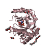
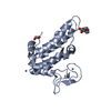
| Deposited unit | 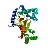
| ||||||||
|---|---|---|---|---|---|---|---|---|---|
| 1 |
| ||||||||
| Unit cell |
|
-Components
| #1: Protein | Mass: 23666.775 Da / Num. of mol.: 1 Source method: isolated from a genetically manipulated source Source: (gene. exp.) STAPHYLOCOCCUS AUREUS (bacteria) / Description: SYNTHETIC GENE / Production host: |
|---|---|
| #2: Water | ChemComp-HOH /  Mass: 18.015 Da / Num. of mol.: 284 / Source method: isolated from a natural source / Formula: H2O Mass: 18.015 Da / Num. of mol.: 284 / Source method: isolated from a natural source / Formula: H2O |
-Experimental details
-Experiment
| Experiment | Method: X-RAY DIFFRACTION / Number of used crystals: 1 |
|---|
- Sample preparation
| Crystal | Density Matthews: 1.75 Å3/Da / Density % sol: 50 % | ||||||||||||||||||||||||
|---|---|---|---|---|---|---|---|---|---|---|---|---|---|---|---|---|---|---|---|---|---|---|---|---|---|
| Crystal grow | pH: 6.5 / Details: 30% PEG 8000, 0.1M SODIUM CACODYLATE BUFFER PH 6.5 | ||||||||||||||||||||||||
| Crystal grow | *PLUS Temperature: 22 ℃ / Method: vapor diffusion, hanging drop / PH range low: 6.6 / PH range high: 6.4 | ||||||||||||||||||||||||
| Components of the solutions | *PLUS
|
-Data collection
| Diffraction | Mean temperature: 100 K |
|---|---|
| Diffraction source | Source: SYNCHROTRON / Site: SRS  / Beamline: PX9.6 / Wavelength: 0.87 / Beamline: PX9.6 / Wavelength: 0.87 |
| Detector | Type: ADSC CCD / Detector: CCD / Date: Dec 22, 2002 |
| Radiation | Monochromator: GRAPHITE / Protocol: SINGLE WAVELENGTH / Monochromatic (M) / Laue (L): M / Scattering type: x-ray |
| Radiation wavelength | Wavelength: 0.87 Å / Relative weight: 1 |
| Reflection | Resolution: 1.68→50 Å / Num. obs: 22478 / % possible obs: 98.9 % / Redundancy: 20 % / Rmerge(I) obs: 0.085 / Net I/σ(I): 20 |
| Reflection shell | Resolution: 1.68→1.74 Å / Rmerge(I) obs: 0.295 / Mean I/σ(I) obs: 5.3 / % possible all: 97.5 |
| Reflection | *PLUS Highest resolution: 1.68 Å / Num. obs: 22619 / % possible obs: 99.4 % / Num. measured all: 409845 / Rmerge(I) obs: 0.085 |
| Reflection shell | *PLUS % possible obs: 97.5 % / Rmerge(I) obs: 0.295 / Mean I/σ(I) obs: 5.3 |
- Processing
| Software |
| |||||||||||||||||||||||||||||||||
|---|---|---|---|---|---|---|---|---|---|---|---|---|---|---|---|---|---|---|---|---|---|---|---|---|---|---|---|---|---|---|---|---|---|---|
| Refinement | Method to determine structure: MOLECULAR REPLACEMENT / Resolution: 1.68→50 Å / Num. parameters: 7863 / Num. restraintsaints: 6859 / Cross valid method: FREE R-VALUE / σ(F): 0 / Stereochemistry target values: ENGH AND HUBER Details: CNS (BRUNGER ET AL) WAS USED IN THE INITIAL STAGES OF REFINEMENT. RESIDUES 197-199 ARE SLIGHTLY DISORDERED AND MODELLED ON THE BASIS OF WEAK DENSITY. THE EXTREMITIES OF SIDE CHAINS 85, ...Details: CNS (BRUNGER ET AL) WAS USED IN THE INITIAL STAGES OF REFINEMENT. RESIDUES 197-199 ARE SLIGHTLY DISORDERED AND MODELLED ON THE BASIS OF WEAK DENSITY. THE EXTREMITIES OF SIDE CHAINS 85, 94,170 AND 200 HAVE BEEN MODELLED AT ZERO OCCUPENCY DUE TO INSUFFICIENT DENSITY.
| |||||||||||||||||||||||||||||||||
| Solvent computation | Solvent model: MOEWS & KRETSINGER, J.MOL.BIOL.91(1973)201-2 | |||||||||||||||||||||||||||||||||
| Refine analyze | Num. disordered residues: 5 / Occupancy sum non hydrogen: 1934.98 | |||||||||||||||||||||||||||||||||
| Refinement step | Cycle: LAST / Resolution: 1.68→50 Å
| |||||||||||||||||||||||||||||||||
| Refine LS restraints |
| |||||||||||||||||||||||||||||||||
| Software | *PLUS Name: SHELXL / Version: 97 / Classification: refinement | |||||||||||||||||||||||||||||||||
| Refinement | *PLUS Rfactor Rwork: 0.17 | |||||||||||||||||||||||||||||||||
| Solvent computation | *PLUS | |||||||||||||||||||||||||||||||||
| Displacement parameters | *PLUS | |||||||||||||||||||||||||||||||||
| Refine LS restraints | *PLUS
|
