 Movie
Movie Controller


+ Open data

- Basic information
| Entry | Database: PDB / ID: 1oft | ||||||
|---|---|---|---|---|---|---|---|
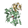
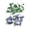
| Title | Crystal structure of SulA from Pseudomonas aeruginosa | ||||||
 Components Components | HYPOTHETICAL PROTEIN PA3008 | ||||||
 Keywords Keywords | BACTERIAL CELL DIVISION INHIBITOR / FTSZ / SULA PROTEIN | ||||||
| Function / homology |  Function and homology information Function and homology informationnegative regulation of cell division / division septum assembly / SOS response / DNA repair Similarity search - Function | ||||||
| Biological species |   PSEUDOMONAS AERUGINOSA (bacteria) PSEUDOMONAS AERUGINOSA (bacteria) | ||||||
| Method |  X-RAY DIFFRACTION / SYNCHROTRON / MIR / Resolution: 2.9 Å X-RAY DIFFRACTION / SYNCHROTRON / MIR / Resolution: 2.9 Å | ||||||
 Authors Authors | Cordell, S.C. / Robinson, E.J.H. / Lowe, J. | ||||||
 Citation Citation | Journal: Proc.Natl.Acad.Sci.USA / Year: 2003 Title: Crystal Structure of the SOS Cell Division Inhibitor Sula and in Complex with Ftsz Authors: Cordell, S.C. / Robinson, E.J.H. / Lowe, J. | ||||||
| History |
|
- Structure visualization
| Structure viewer | Molecule: MolmilJmol/JSmol |
|---|
- Downloads & links
-Download
| PDBx/mmCIF format | 1oft.cif.gz | 99.5 KB | Display | PDBx/mmCIF format |
|---|---|---|---|---|
| PDB format | pdb1oft.ent.gz | 78.2 KB | Display | PDB format |
| PDBx/mmJSON format | 1oft.json.gz | Tree view | PDBx/mmJSON format | |
| Others |  Other downloads Other downloads |
-Validation report
| Arichive directory | https://data.pdbj.org/pub/pdb/validation_reports/of/1oftftp://data.pdbj.org/pub/pdb/validation_reports/of/1oft | HTTPS FTP |
|---|
-Related structure data


-Links
 PDBj
PDBj- Assembly
| Deposited unit | 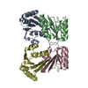
| ||||||||
|---|---|---|---|---|---|---|---|---|---|
| 1 |
| ||||||||
| Unit cell |
|
-Components
| #1: Protein | Mass: 17493.922 Da / Num. of mol.: 4 Source method: isolated from a genetically manipulated source Source: (gene. exp.) PSEUDOMONAS AERUGINOSA (bacteria) / Plasmid: PHIS17 / Production host: |
|---|
-Experimental details
-Experiment
| Experiment | Method: X-RAY DIFFRACTION / Number of used crystals: 1 |
|---|
- Sample preparation
| Crystal | Density Matthews: 2.51 Å3/Da / Density % sol: 50.96 % | ||||||||||||||||||||||||||||||||||||||||||||||||||||||||||||||||||||||||||||||||||||
|---|---|---|---|---|---|---|---|---|---|---|---|---|---|---|---|---|---|---|---|---|---|---|---|---|---|---|---|---|---|---|---|---|---|---|---|---|---|---|---|---|---|---|---|---|---|---|---|---|---|---|---|---|---|---|---|---|---|---|---|---|---|---|---|---|---|---|---|---|---|---|---|---|---|---|---|---|---|---|---|---|---|---|---|---|---|
| Crystal grow | pH: 5.6 / Details: pH 5.60 | ||||||||||||||||||||||||||||||||||||||||||||||||||||||||||||||||||||||||||||||||||||
| Crystal grow | *PLUS Temperature: 19 ℃ / pH: 7.5 / Method: vapor diffusion, sitting drop | ||||||||||||||||||||||||||||||||||||||||||||||||||||||||||||||||||||||||||||||||||||
| Components of the solutions | *PLUS
|
-Data collection
| Diffraction | Mean temperature: 100 K |
|---|---|
| Diffraction source | Source: SYNCHROTRON / Site: SRS  / Beamline: PX9.6 / Wavelength: 0.8638 / Beamline: PX9.6 / Wavelength: 0.8638 |
| Detector | Type: ADSC CCD / Detector: CCD |
| Radiation | Protocol: SINGLE WAVELENGTH / Monochromatic (M) / Laue (L): M / Scattering type: x-ray |
| Radiation wavelength | Wavelength: 0.8638 Å / Relative weight: 1 |
| Reflection | Resolution: 2.9→50 Å / Num. obs: 15552 / % possible obs: 96.3 % / Redundancy: 3.3 % / Biso Wilson estimate: 96 Å2 / Rmerge(I) obs: 0.075 / Net I/σ(I): 14 |
| Reflection shell | Resolution: 2.9→3.06 Å / Redundancy: 2.9 % / Rmerge(I) obs: 0.391 / Mean I/σ(I) obs: 2.2 / % possible all: 96.3 |
| Reflection | *PLUS Highest resolution: 2.9 Å / % possible obs: 99.8 % / Redundancy: 3.9 % / Rmerge(I) obs: 0.095 |
| Reflection shell | *PLUS % possible obs: 99.8 % / Rmerge(I) obs: 0.274 / Mean I/σ(I) obs: 4.4 |
- Processing
| Software |
| ||||||||||||||||||||||||||||||||||||||||||||||||||||||||||||
|---|---|---|---|---|---|---|---|---|---|---|---|---|---|---|---|---|---|---|---|---|---|---|---|---|---|---|---|---|---|---|---|---|---|---|---|---|---|---|---|---|---|---|---|---|---|---|---|---|---|---|---|---|---|---|---|---|---|---|---|---|---|
| Refinement | Method to determine structure: MIR / Resolution: 2.9→50 Å / Isotropic thermal model: RESTRAINED / Cross valid method: THROUGHOUT / σ(F): 0 / Details: DATA SHARPENED BY B= -40A**2
| ||||||||||||||||||||||||||||||||||||||||||||||||||||||||||||
| Displacement parameters | Biso mean: 35 Å2
| ||||||||||||||||||||||||||||||||||||||||||||||||||||||||||||
| Refinement step | Cycle: LAST / Resolution: 2.9→50 Å
| ||||||||||||||||||||||||||||||||||||||||||||||||||||||||||||
| Refine LS restraints |
| ||||||||||||||||||||||||||||||||||||||||||||||||||||||||||||
| Refine LS restraints NCS | NCS model details: RESTRAINTS | ||||||||||||||||||||||||||||||||||||||||||||||||||||||||||||
| LS refinement shell | Resolution: 2.9→2.97 Å / Total num. of bins used: 15
| ||||||||||||||||||||||||||||||||||||||||||||||||||||||||||||
| Refinement | *PLUS Rfactor Rfree: 0.289 / Rfactor Rwork: 0.247 | ||||||||||||||||||||||||||||||||||||||||||||||||||||||||||||
| Solvent computation | *PLUS | ||||||||||||||||||||||||||||||||||||||||||||||||||||||||||||
| Displacement parameters | *PLUS | ||||||||||||||||||||||||||||||||||||||||||||||||||||||||||||
| LS refinement shell | *PLUS Rfactor Rfree: 0.366 / Rfactor Rwork: 0.258 |
