 Movie
Movie Controller


+ Open data

- Basic information
| Entry | Database: PDB / ID: 1ofu | ||||||
|---|---|---|---|---|---|---|---|
| Title | Crystal structure of SulA:FtsZ from Pseudomonas aeruginosa | ||||||
 Components Components |
| ||||||
 Keywords Keywords | BACTERIAL CELL DIVISION INHIBITOR / FTSZ / SULA PROTEIN | ||||||
| Function / homology |  Function and homology information Function and homology informationnegative regulation of cell division / division septum assembly / FtsZ-dependent cytokinesis / SOS response / cell division site / protein polymerization / cell division / DNA repair / GTPase activity / GTP binding / cytoplasm Similarity search - Function | ||||||
| Biological species |   PSEUDOMONAS AERUGINOSA (bacteria) PSEUDOMONAS AERUGINOSA (bacteria) | ||||||
| Method |  X-RAY DIFFRACTION / SYNCHROTRON / MOLECULAR REPLACEMENT / Resolution: 2.1 Å X-RAY DIFFRACTION / SYNCHROTRON / MOLECULAR REPLACEMENT / Resolution: 2.1 Å | ||||||
 Authors Authors | Cordell, S.C. / Robinson, E.J.H. / Lowe, J. | ||||||
 Citation Citation | Journal: Proc.Natl.Acad.Sci.USA / Year: 2003 Title: Crystal Structure of the SOS Cell Division Inhibitor Sula and in Complex with Ftsz Authors: Cordell, S.C. / Robinson, E.J.H. / Lowe, J. | ||||||
| History |
|
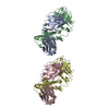
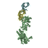
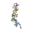
- Structure visualization
| Structure viewer | Molecule: MolmilJmol/JSmol |
|---|
- Downloads & links
-Download
| PDBx/mmCIF format | 1ofu.cif.gz | 175.9 KB | Display | PDBx/mmCIF format |
|---|---|---|---|---|
| PDB format | pdb1ofu.ent.gz | 140.2 KB | Display | PDB format |
| PDBx/mmJSON format | 1ofu.json.gz | Tree view | PDBx/mmJSON format | |
| Others |  Other downloads Other downloads |
-Validation report
| Arichive directory | https://data.pdbj.org/pub/pdb/validation_reports/of/1ofuftp://data.pdbj.org/pub/pdb/validation_reports/of/1ofu | HTTPS FTP |
|---|
-Related structure data


-Links
 PDBj
PDBj


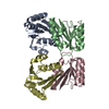
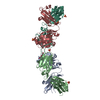
- Assembly
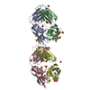
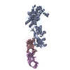
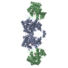
| Deposited unit | 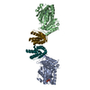
| ||||||||
|---|---|---|---|---|---|---|---|---|---|
| 1 |
| ||||||||
| Unit cell |
|
-Components
| #1: Protein | Mass: 33146.961 Da / Num. of mol.: 2 / Fragment: RESIDUES 1-320 Source method: isolated from a genetically manipulated source Details: LAST 75 RESIDUES REMOVED FROM C-TERMINUS / Source: (gene. exp.) PSEUDOMONAS AERUGINOSA (bacteria) / Plasmid: PHIS17 / Production host: #2: Protein | Mass: 13023.056 Da / Num. of mol.: 2 / Fragment: RESIDUES 43-161 Source method: isolated from a genetically manipulated source Details: FIRST 42 RESIDUES REMOVED FROM N-TERMINUS / Source: (gene. exp.) PSEUDOMONAS AERUGINOSA (bacteria) / Plasmid: PHIS17 / Production host: #3: Chemical |   Type: RNA linking / Mass: 443.201 Da / Num. of mol.: 2 / Source method: obtained synthetically / Formula: C10H15N5O11P2 / Comment: GDP, energy-carrying molecule*YM Type: RNA linking / Mass: 443.201 Da / Num. of mol.: 2 / Source method: obtained synthetically / Formula: C10H15N5O11P2 / Comment: GDP, energy-carrying molecule*YM#4: Water | ChemComp-HOH / |  Mass: 18.015 Da / Num. of mol.: 441 / Source method: isolated from a natural source / Formula: H2O Mass: 18.015 Da / Num. of mol.: 441 / Source method: isolated from a natural source / Formula: H2O |
|---|
-Experimental details
-Experiment
| Experiment | Method: X-RAY DIFFRACTION / Number of used crystals: 1 |
|---|
- Sample preparation
| Crystal | Density Matthews: 2.74 Å3/Da / Density % sol: 55.04 % | |||||||||||||||||||||||||||||||||||||||||||||||||||||||||||||||||||||||||||||
|---|---|---|---|---|---|---|---|---|---|---|---|---|---|---|---|---|---|---|---|---|---|---|---|---|---|---|---|---|---|---|---|---|---|---|---|---|---|---|---|---|---|---|---|---|---|---|---|---|---|---|---|---|---|---|---|---|---|---|---|---|---|---|---|---|---|---|---|---|---|---|---|---|---|---|---|---|---|---|
| Crystal grow | pH: 5.6 / Details: pH 5.60 | |||||||||||||||||||||||||||||||||||||||||||||||||||||||||||||||||||||||||||||
| Crystal grow | *PLUS Temperature: 19 ℃ / pH: 7.5 / Method: vapor diffusion, sitting drop | |||||||||||||||||||||||||||||||||||||||||||||||||||||||||||||||||||||||||||||
| Components of the solutions | *PLUS
|
-Data collection
| Diffraction | Mean temperature: 100 K |
|---|---|
| Diffraction source | Source: SYNCHROTRON / Site: ESRF  / Beamline: ID14-4 / Wavelength: 0.9393 / Beamline: ID14-4 / Wavelength: 0.9393 |
| Detector | Detector: CCD |
| Radiation | Protocol: SINGLE WAVELENGTH / Monochromatic (M) / Laue (L): M / Scattering type: x-ray |
| Radiation wavelength | Wavelength: 0.9393 Å / Relative weight: 1 |
| Reflection | Resolution: 2.1→50 Å / Num. obs: 60044 / % possible obs: 99.7 % / Redundancy: 3.7 % / Biso Wilson estimate: 34.8 Å2 / Rmerge(I) obs: 0.096 / Net I/σ(I): 11.4 |
| Reflection shell | Resolution: 2.1→2.21 Å / Redundancy: 3.8 % / Rmerge(I) obs: 0.334 / Mean I/σ(I) obs: 3.7 / % possible all: 99.7 |
| Reflection | *PLUS Highest resolution: 2.1 Å / Redundancy: 3.8 % / Rmerge(I) obs: 0.096 |
| Reflection shell | *PLUS % possible obs: 99.7 % / Rmerge(I) obs: 0.334 / Mean I/σ(I) obs: 3.7 |
- Processing
| Software |
| ||||||||||||||||||||||||||||||||||||||||||||||||||||||||||||
|---|---|---|---|---|---|---|---|---|---|---|---|---|---|---|---|---|---|---|---|---|---|---|---|---|---|---|---|---|---|---|---|---|---|---|---|---|---|---|---|---|---|---|---|---|---|---|---|---|---|---|---|---|---|---|---|---|---|---|---|---|---|
| Refinement | Method to determine structure: MOLECULAR REPLACEMENT Starting model: MAD MODEL FROM C2 CRYSTAL FORM Resolution: 2.1→50 Å / Isotropic thermal model: RESTRAINED / Cross valid method: THROUGHOUT / σ(F): 0
| ||||||||||||||||||||||||||||||||||||||||||||||||||||||||||||
| Displacement parameters | Biso mean: 43.7 Å2
| ||||||||||||||||||||||||||||||||||||||||||||||||||||||||||||
| Refinement step | Cycle: LAST / Resolution: 2.1→50 Å
| ||||||||||||||||||||||||||||||||||||||||||||||||||||||||||||
| Refine LS restraints |
| ||||||||||||||||||||||||||||||||||||||||||||||||||||||||||||
| Refine LS restraints NCS | NCS model details: RESTRAINTS | ||||||||||||||||||||||||||||||||||||||||||||||||||||||||||||
| LS refinement shell | Resolution: 2.1→2.11 Å / Total num. of bins used: 50
| ||||||||||||||||||||||||||||||||||||||||||||||||||||||||||||
| Refinement | *PLUS Rfactor Rfree: 0.255 / Rfactor Rwork: 0.216 | ||||||||||||||||||||||||||||||||||||||||||||||||||||||||||||
| Solvent computation | *PLUS | ||||||||||||||||||||||||||||||||||||||||||||||||||||||||||||
| Displacement parameters | *PLUS | ||||||||||||||||||||||||||||||||||||||||||||||||||||||||||||
| LS refinement shell | *PLUS Rfactor Rfree: 0.405 / Rfactor Rwork: 0.354 |
