 Movie
Movie Controller


+ Open data

- Basic information
| Entry | Database: PDB / ID: 6wvg | |||||||||||||||||||||
|---|---|---|---|---|---|---|---|---|---|---|---|---|---|---|---|---|---|---|---|---|---|---|
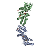
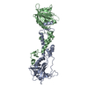
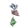
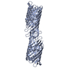
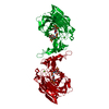
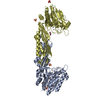
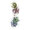
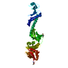
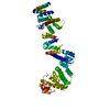
| Title | human CD53 | |||||||||||||||||||||
 Components Components | Green fluorescent protein, Leukocyte surface antigen CD53 chimera | |||||||||||||||||||||
 Keywords Keywords | MEMBRANE PROTEIN / CD53 / tetraspanin-25 | |||||||||||||||||||||
| Function / homology |  Function and homology information Function and homology informationpositive regulation of myoblast fusion / receptor clustering / tertiary granule membrane / immunological synapse / specific granule membrane / protein-membrane adaptor activity / bioluminescence / generation of precursor metabolites and energy / cell-cell junction / Neutrophil degranulation ...positive regulation of myoblast fusion / receptor clustering / tertiary granule membrane / immunological synapse / specific granule membrane / protein-membrane adaptor activity / bioluminescence / generation of precursor metabolites and energy / cell-cell junction / Neutrophil degranulation / synapse / cell surface / signal transduction / extracellular exosome / identical protein binding / plasma membrane Similarity search - Function | |||||||||||||||||||||
| Biological species |   Aequorea victoria (jellyfish) Aequorea victoria (jellyfish) Homo sapiens (human) Homo sapiens (human) | |||||||||||||||||||||
| Method |  X-RAY DIFFRACTION / SYNCHROTRON / MOLECULAR REPLACEMENT / Resolution: 2.9 Å X-RAY DIFFRACTION / SYNCHROTRON / MOLECULAR REPLACEMENT / Resolution: 2.9 Å | |||||||||||||||||||||
 Authors Authors | Yang, Y. / Liu, S. / Li, W. | |||||||||||||||||||||
| Funding support |  United States, 6items United States, 6items
| |||||||||||||||||||||
 Citation Citation | Journal: Embo J. / Year: 2020 Title: Open conformation of tetraspanins shapes interaction partner networks on cell membranes. Authors: Yang, Y. / Liu, X.R. / Greenberg, Z.J. / Zhou, F. / He, P. / Fan, L. / Liu, S. / Shen, G. / Egawa, T. / Gross, M.L. / Schuettpelz, L.G. / Li, W. #1: Journal: To Be PublishedTitle: Termini restraining of small membrane proteins enables structure determination at atomic resolution Authors: Liu, S. / Li, S. / Yang, Y. / Li, W. | |||||||||||||||||||||
| History |
|
- Structure visualization
| Structure viewer | Molecule: MolmilJmol/JSmol |
|---|
- Downloads & links
-Download
| PDBx/mmCIF format | 6wvg.cif.gz | 365 KB | Display | PDBx/mmCIF format |
|---|---|---|---|---|
| PDB format | pdb6wvg.ent.gz | 297 KB | Display | PDB format |
| PDBx/mmJSON format | 6wvg.json.gz | Tree view | PDBx/mmJSON format | |
| Others |  Other downloads Other downloads |
-Validation report
| Arichive directory | https://data.pdbj.org/pub/pdb/validation_reports/wv/6wvgftp://data.pdbj.org/pub/pdb/validation_reports/wv/6wvg | HTTPS FTP |
|---|
-Related structure data
| Related structure data |  2b3pS S: Starting model for refinement |
|---|---|
| Similar structure data |

-Links
 PDBj
PDBj


- Assembly
| Deposited unit | 
| |||||||||||||||||||||||||||||||||||||||||||||||||||||||||||||||||||||||||||||||||||||||||||||||||||||||||||||||||
|---|---|---|---|---|---|---|---|---|---|---|---|---|---|---|---|---|---|---|---|---|---|---|---|---|---|---|---|---|---|---|---|---|---|---|---|---|---|---|---|---|---|---|---|---|---|---|---|---|---|---|---|---|---|---|---|---|---|---|---|---|---|---|---|---|---|---|---|---|---|---|---|---|---|---|---|---|---|---|---|---|---|---|---|---|---|---|---|---|---|---|---|---|---|---|---|---|---|---|---|---|---|---|---|---|---|---|---|---|---|---|---|---|---|---|
| 1 | 
| |||||||||||||||||||||||||||||||||||||||||||||||||||||||||||||||||||||||||||||||||||||||||||||||||||||||||||||||||
| 2 | 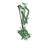
| |||||||||||||||||||||||||||||||||||||||||||||||||||||||||||||||||||||||||||||||||||||||||||||||||||||||||||||||||
| Unit cell |
| |||||||||||||||||||||||||||||||||||||||||||||||||||||||||||||||||||||||||||||||||||||||||||||||||||||||||||||||||
| Noncrystallographic symmetry (NCS) | NCS domain:
NCS domain segments:
|