 Movie
Movie Controller

[English] 日本語
 Yorodumi
- PDB-5svv: Structure and kinetics of the LOV domain of ZEITLUPE determine it... -
+ Open data

- Basic information
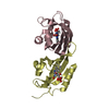
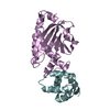
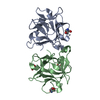
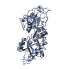
| Entry | Database: PDB / ID: 5svv | ||||||
|---|---|---|---|---|---|---|---|
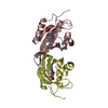
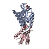
| Title | Structure and kinetics of the LOV domain of ZEITLUPE determine its circadian function in Arabidopsis | ||||||
 Components Components | Adagio protein 1 | ||||||
 Keywords Keywords | CIRCADIAN CLOCK PROTEIN / LOV / Kinetics / PAS domain / photoreceptor | ||||||
| Function / homology |  Function and homology information Function and homology informationresponse to red light / flower development / blue light photoreceptor activity / response to blue light / SCF ubiquitin ligase complex / entrainment of circadian clock by photoperiod / SCF-dependent proteasomal ubiquitin-dependent protein catabolic process / proteasomal protein catabolic process / circadian rhythm / regulation of circadian rhythm ...response to red light / flower development / blue light photoreceptor activity / response to blue light / SCF ubiquitin ligase complex / entrainment of circadian clock by photoperiod / SCF-dependent proteasomal ubiquitin-dependent protein catabolic process / proteasomal protein catabolic process / circadian rhythm / regulation of circadian rhythm / ubiquitin-dependent protein catabolic process / protein ubiquitination / nucleus / cytosol Similarity search - Function | ||||||
| Biological species |  | ||||||
| Method |  X-RAY DIFFRACTION / SYNCHROTRON / MOLECULAR REPLACEMENT / Resolution: 2.101 Å X-RAY DIFFRACTION / SYNCHROTRON / MOLECULAR REPLACEMENT / Resolution: 2.101 Å | ||||||
 Authors Authors | Zoltowski, B. / Pudasaini, A. | ||||||
| Funding support |  United States, 1items United States, 1items
| ||||||
 Citation Citation | Journal: Elife / Year: 2017 Title: Kinetics of the LOV domain of ZEITLUPE determine its circadian function inArabidopsis. Authors: Pudasaini, A. / Shim, J.S. / Song, Y.H. / Shi, H. / Kiba, T. / Somers, D.E. / Imaizumi, T. / Zoltowski, B.D. | ||||||
| History |
|
- Structure visualization
| Structure viewer | Molecule: MolmilJmol/JSmol |
|---|
- Downloads & links
-Download
| PDBx/mmCIF format | 5svv.cif.gz | 202.9 KB | Display | PDBx/mmCIF format |
|---|---|---|---|---|
| PDB format | pdb5svv.ent.gz | 165.1 KB | Display | PDB format |
| PDBx/mmJSON format | 5svv.json.gz | Tree view | PDBx/mmJSON format | |
| Others |  Other downloads Other downloads |
-Validation report
| Arichive directory | https://data.pdbj.org/pub/pdb/validation_reports/sv/5svvftp://data.pdbj.org/pub/pdb/validation_reports/sv/5svv | HTTPS FTP |
|---|
-Related structure data
| Related structure data |  5svgC 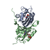 5svuC 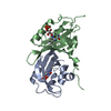 5svwC 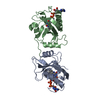 3d72S C: citing same article ( S: Starting model for refinement |
|---|---|
| Similar structure data |


-Links
 PDBj
PDBj


- Assembly
| Deposited unit | 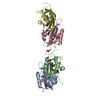
| ||||||||
|---|---|---|---|---|---|---|---|---|---|
| 1 | 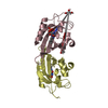
| ||||||||
| 2 | 
| ||||||||
| Unit cell |
|
-Components
| #1: Protein | Mass: 15408.677 Da / Num. of mol.: 4 / Fragment: LOV domain (UNP residues 29-165) / Mutation: V48I, G80R Source method: isolated from a genetically manipulated source Source: (gene. exp.)  #2: Chemical | ChemComp-FMN /   Mass: 456.344 Da / Num. of mol.: 4 / Mutation: V48I, G80R Mass: 456.344 Da / Num. of mol.: 4 / Mutation: V48I, G80RSource method: isolated from a genetically manipulated source Formula: C17H21N4O9P #3: Chemical |   Mass: 59.044 Da / Num. of mol.: 3 / Source method: obtained synthetically / Formula: C2H3O2 Mass: 59.044 Da / Num. of mol.: 3 / Source method: obtained synthetically / Formula: C2H3O2#4: Chemical |   Mass: 92.094 Da / Num. of mol.: 2 / Source method: obtained synthetically / Formula: C3H8O3 Mass: 92.094 Da / Num. of mol.: 2 / Source method: obtained synthetically / Formula: C3H8O3#5: Water | ChemComp-HOH / |  Mass: 18.015 Da / Num. of mol.: 343 / Source method: isolated from a natural source / Formula: H2O Mass: 18.015 Da / Num. of mol.: 343 / Source method: isolated from a natural source / Formula: H2O |
|---|
-Experimental details
-Experiment
| Experiment | Method: X-RAY DIFFRACTION / Number of used crystals: 1 |
|---|
- Sample preparation
| Crystal | Density Matthews: 3.41 Å3/Da / Density % sol: 63.97 % |
|---|---|
| Crystal grow | Temperature: 298 K / Method: vapor diffusion, hanging drop Details: 0.1 M Tris ph 8.5, 0.2 M Sodium Acetate trihydrate, 30% w/v PEG 4K |
-Data collection
| Diffraction | Mean temperature: 80 K |
|---|---|
| Diffraction source | Source: SYNCHROTRON / Site: CHESS / Beamline: F1 / Wavelength: 0.979 Å |
| Detector | Type: ADSC QUANTUM 270 / Detector: CCD / Date: Mar 6, 2014 |
| Radiation | Protocol: SINGLE WAVELENGTH / Monochromatic (M) / Laue (L): M / Scattering type: x-ray |
| Radiation wavelength | Wavelength: 0.979 Å / Relative weight: 1 |
| Reflection | Resolution: 2.1→50 Å / Num. obs: 49510 / % possible obs: 99.3 % / Redundancy: 7.9 % / Net I/σ(I): 36.6 |
| Reflection shell | Resolution: 2.1→2.14 Å / Redundancy: 8.2 % / Rmerge(I) obs: 0.251 / Mean I/σ(I) obs: 12.8 / % possible all: 100 |
- Processing
| Software |
| |||||||||||||||||||||||||||||||||||||||||||||||||||||||||||||||||||||||||||||||||||||||||||||||||||||||||||||||||||||||||||||||||||||
|---|---|---|---|---|---|---|---|---|---|---|---|---|---|---|---|---|---|---|---|---|---|---|---|---|---|---|---|---|---|---|---|---|---|---|---|---|---|---|---|---|---|---|---|---|---|---|---|---|---|---|---|---|---|---|---|---|---|---|---|---|---|---|---|---|---|---|---|---|---|---|---|---|---|---|---|---|---|---|---|---|---|---|---|---|---|---|---|---|---|---|---|---|---|---|---|---|---|---|---|---|---|---|---|---|---|---|---|---|---|---|---|---|---|---|---|---|---|---|---|---|---|---|---|---|---|---|---|---|---|---|---|---|---|---|
| Refinement | Method to determine structure: MOLECULAR REPLACEMENT Starting model: 3D72 Resolution: 2.101→36.367 Å / SU ML: 0.17 / Cross valid method: FREE R-VALUE / σ(F): 1.34 / Phase error: 18.29 / Stereochemistry target values: ML
| |||||||||||||||||||||||||||||||||||||||||||||||||||||||||||||||||||||||||||||||||||||||||||||||||||||||||||||||||||||||||||||||||||||
| Solvent computation | Shrinkage radii: 0.9 Å / VDW probe radii: 1.11 Å / Solvent model: FLAT BULK SOLVENT MODEL | |||||||||||||||||||||||||||||||||||||||||||||||||||||||||||||||||||||||||||||||||||||||||||||||||||||||||||||||||||||||||||||||||||||
| Refinement step | Cycle: LAST / Resolution: 2.101→36.367 Å
| |||||||||||||||||||||||||||||||||||||||||||||||||||||||||||||||||||||||||||||||||||||||||||||||||||||||||||||||||||||||||||||||||||||
| Refine LS restraints |
| |||||||||||||||||||||||||||||||||||||||||||||||||||||||||||||||||||||||||||||||||||||||||||||||||||||||||||||||||||||||||||||||||||||
| LS refinement shell |
|
