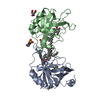
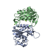
SHEET THE SHEET STRUCTURE OF THIS MOLECULE IS BIFURCATED. IN ORDER TO REPRESENT THIS FEATURE IN ... SHEET THE SHEET STRUCTURE OF THIS MOLECULE IS BIFURCATED. IN ORDER TO REPRESENT THIS FEATURE IN THE SHEET RECORDS BELOW, TWO SHEETS ARE DEFINED.
Monochromator: BM14 / Protocol: SINGLE WAVELENGTH / Monochromatic (M) / Laue (L): M / Scattering type: x-ray
Radiation wavelength
Wavelength: 0.7513 Å / Relative weight: 1
Reflection
Resolution: 2→65.94 Å / Num. obs: 26054 / % possible obs: 99.2 %
Reflection
*PLUS
Highest resolution: 2 Å / Lowest resolution: 65.94 Å
-
Processing
Software
Name
Classification
REFMAC
refinement
DENZO
datareduction
SCALEPACK
datascaling
Refinement
Method to determine structure: OTHER / Resolution: 2→65.94 Å / SU B: 5.6 / SU ML: 0.159 / Cross valid method: THROUGHOUT / ESU R: 0.176 / ESU R Free: 0.163
Rfactor
Num. reflection
% reflection
Selection details
Rfree
0.227
1326
5.1 %
RANDOM
Rwork
0.174
-
-
-
obs
0.177
24728
99.19 %
-
Displacement parameters
Biso mean: 19 Å2
Baniso -1
Baniso -2
Baniso -3
1-
0.54 Å2
0 Å2
0 Å2
2-
-
0.23 Å2
0 Å2
3-
-
-
-0.77 Å2
Refinement step
Cycle: LAST / Resolution: 2→65.94 Å
Protein
Nucleic acid
Ligand
Solvent
Total
Num. atoms
2913
0
0
146
3059
Refinement
*PLUS
Highest resolution: 2 Å
Solvent computation
*PLUS
Displacement parameters
*PLUS
Refine LS restraints
*PLUS
Refine-ID
Type
Dev ideal
X-RAY DIFFRACTION
bond_d
0.034
X-RAY DIFFRACTION
angle_d
X-RAY DIFFRACTION
angle_deg
2.5
X-RAY DIFFRACTION
dihedral_angle_d
X-RAY DIFFRACTION
dihedral_angle_deg
5.7
+
About Yorodumi
-
News
-
Feb 9, 2022. New format data for meta-information of EMDB entries
New format data for meta-information of EMDB entries
Version 3 of the EMDB header file is now the official format.
The previous official version 1.9 will be removed from the archive.
In the structure databanks used in Yorodumi, some data are registered as the other names, "COVID-19 virus" and "2019-nCoV". Here are the details of the virus and the list of structure data.
Jan 31, 2019. EMDB accession codes are about to change! (news from PDBe EMDB page)
EMDB accession codes are about to change! (news from PDBe EMDB page)
The allocation of 4 digits for EMDB accession codes will soon come to an end. Whilst these codes will remain in use, new EMDB accession codes will include an additional digit and will expand incrementally as the available range of codes is exhausted. The current 4-digit format prefixed with “EMD-” (i.e. EMD-XXXX) will advance to a 5-digit format (i.e. EMD-XXXXX), and so on. It is currently estimated that the 4-digit codes will be depleted around Spring 2019, at which point the 5-digit format will come into force.
The EM Navigator/Yorodumi systems omit the EMD- prefix.
Related info.:Q: What is EMD? / ID/Accession-code notation in Yorodumi/EM Navigator
Yorodumi is a browser for structure data from EMDB, PDB, SASBDB, etc.
This page is also the successor to EM Navigator detail page, and also detail information page/front-end page for Omokage search.
The word "yorodu" (or yorozu) is an old Japanese word meaning "ten thousand". "mi" (miru) is to see.
Related info.:EMDB / PDB / SASBDB / Comparison of 3 databanks / Yorodumi Search / Aug 31, 2016. New EM Navigator & Yorodumi / Yorodumi Papers / Jmol/JSmol / Function and homology information / Changes in new EM Navigator and Yorodumi
 Movie
Movie Controller

 Yorodumi
 Open data
 Basic information
 Components
 Keywords
 Function and homology information
 HOMARUS GAMMARUS (European lobster)
 X-RAY DIFFRACTION /
  Authors
 Citation
 Structure visualization
 Downloads & links
 Other downloads


 PDBj
PDBj

 Assembly
 Mass: 18.015 Da / Num. of mol.: 146 / Source method: isolated from a natural source / Formula: H2O
 Sample preparation
 / Beamline: BM14 / Wavelength: 0.7513
  Processing