 Movie
Movie Controller

[English] 日本語
 Yorodumi
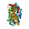
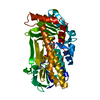
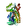
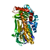
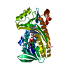
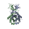
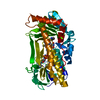
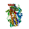
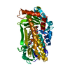
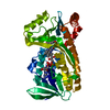
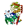
- PDB-1cc6: PHE161 AND ARG166 VARIANTS OF P-HYDROXYBENZOATE HYDROXYLASE. IMPL... -
+ Open data

- Basic information
| Entry | Database: PDB / ID: 1cc6 | ||||||
|---|---|---|---|---|---|---|---|
| Title | PHE161 AND ARG166 VARIANTS OF P-HYDROXYBENZOATE HYDROXYLASE. IMPLICATIONS FOR NADPH RECOGNITION AND STRUCTURAL STABILITY. | ||||||
 Components Components | PROTEIN (P-HYDROXYBENZOATE HYDROXYLASE) | ||||||
 Keywords Keywords | OXIDOREDUCTASE / HYDROXYBENZOATE | ||||||
| Function / homology |  Function and homology information Function and homology information4-hydroxybenzoate 3-monooxygenase (NADPH) activity / 4-hydroxybenzoate 3-monooxygenase / 4-hydroxybenzoate 3-monooxygenase activity / : / FAD binding / flavin adenine dinucleotide binding Similarity search - Function | ||||||
| Biological species |  Pseudomonas fluorescens (bacteria) Pseudomonas fluorescens (bacteria) | ||||||
| Method |  X-RAY DIFFRACTION / MOLECULAR REPLACEMENT / Resolution: 2.2 Å X-RAY DIFFRACTION / MOLECULAR REPLACEMENT / Resolution: 2.2 Å | ||||||
 Authors Authors | Eppink, M.H.M. / Bunthof, C. / Schreuder, H.A. / Van Berkel, W.J.H. | ||||||
 Citation Citation | Journal: Febs Lett. / Year: 1999 Title: Phe161 and Arg166 variants of p-hydroxybenzoate hydroxylase. Implications for NADPH recognition and structural stability. Authors: Eppink, M.H. / Bunthol, C. / Schreuder, H.A. / van Berkel, W.J. #1: Journal: Protein Sci. / Year: 1994Title: Crystal structure of p-hydroxybenzoate hydroxylase reconstituted with the modified FAD present in alcohol oxidase from methylotrophic yeasts: evidence for an arabinoflavin. Authors: van Berkel, W.J. / Eppink, M.H. / Schreuder, H.A. #2: Journal: Biochemistry / Year: 1994Title: Crystal structures of wild-type p-hydroxybenzoate hydroxylase complexed with 4-aminobenzoate,2,4-dihydroxybenzoate, and 2-hydroxy-4-aminobenzoate and of the Tyr222Ala mutant complexed with 2- ...Title: Crystal structures of wild-type p-hydroxybenzoate hydroxylase complexed with 4-aminobenzoate,2,4-dihydroxybenzoate, and 2-hydroxy-4-aminobenzoate and of the Tyr222Ala mutant complexed with 2-hydroxy-4-aminobenzoate. Evidence for a proton channel and a new binding mode of the flavin ring. Authors: Schreuder, H.A. / Mattevi, A. / Obmolova, G. / Kalk, K.H. / Hol, W.G. / van der Bolt, F.J. / van Berkel, W.J. #3: Journal: Proteins / Year: 1992 Title: Crystal structure of the reduced form of p-hydroxybenzoate hydroxylase refined at 2.3 A resolution. Authors: Schreuder, H.A. / van der Laan, J.M. / Swarte, M.B. / Kalk, K.H. / Hol, W.G. / Drenth, J. #4: Journal: Febs Lett. / Year: 1990 Title: Engineering of microheterogeneity-resistant p-hydroxybenzoate hydroxylase from Pseudomonas fluorescens. Authors: Eschrich, K. / van Berkel, W.J. / Westphal, A.H. / de Kok, A. / Mattevi, A. / Obmolova, G. / Kalk, K.H. / Hol, W.G. #5: Journal: J.Mol.Biol. / Year: 1989Title: Crystal structure of the p-hydroxybenzoate hydroxylase-substrate complex refined at 1.9 A resolution. Analysis of the enzyme-substrate and enzyme-product complexes. Authors: Schreuder, H.A. / Prick, P.A. / Wierenga, R.K. / Vriend, G. / Wilson, K.S. / Hol, W.G. / Drenth, J. #6: Journal: Biochemistry / Year: 1989Title: The coenzyme analogue adenosine 5-diphosphoribose displaces FAD in the active site of p-hydroxybenzoate hydroxylase. An x-ray crystallographic investigation. Authors: van der Laan, J.M. / Schreuder, H.A. / Swarte, M.B. / Wierenga, R.K. / Kalk, K.H. / Hol, W.G. / Drenth, J. #7: Journal: J.Mol.Biol. / Year: 1988Title: Crystal structure of p-hydroxybenzoate hydroxylase complexed with its reaction product 3,4-dihydroxybenzoate. Authors: Schreuder, H.A. / van der Laan, J.M. / Hol, W.G. / Drenth, J. #8: Journal: J.Mol.Biol. / Year: 1979 Title: Crystal structure of p-hydroxybenzoate hydroxylase. Authors: Wierenga, R.K. / de Jong, R.J. / Kalk, K.H. / Hol, W.G. / Drenth, J. #9: Journal: J.Biol.Chem. / Year: 1975 Title: Crystallization and preliminary x-ray investigation of p-hydroxybenzoate hydroxylase from Pseudomonas fluorescens. Authors: Drenth, J. / Hol, W.G. / Wierenga, R.K. | ||||||
| History |
|
- Structure visualization
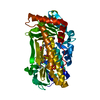
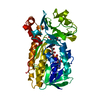
| Structure viewer | Molecule: MolmilJmol/JSmol |
|---|
- Downloads & links
-Download
| PDBx/mmCIF format | 1cc6.cif.gz | 99.6 KB | Display | PDBx/mmCIF format |
|---|---|---|---|---|
| PDB format | pdb1cc6.ent.gz | 74.1 KB | Display | PDB format |
| PDBx/mmJSON format | 1cc6.json.gz | Tree view | PDBx/mmJSON format | |
| Others |  Other downloads Other downloads |
-Validation report
| Arichive directory | https://data.pdbj.org/pub/pdb/validation_reports/cc/1cc6ftp://data.pdbj.org/pub/pdb/validation_reports/cc/1cc6 | HTTPS FTP |
|---|
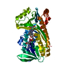
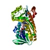
-Related structure data
| Related structure data |  1cc4C  1pbeS S: Starting model for refinement C: citing same article ( |
|---|---|
| Similar structure data |


-Links
 PDBj
PDBj- Assembly
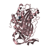
| Deposited unit | 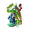
| ||||||||||
|---|---|---|---|---|---|---|---|---|---|---|---|
| 1 |
| ||||||||||
| Unit cell |
|
-Components
| #1: Protein | Mass: 44294.395 Da / Num. of mol.: 1 / Mutation: R166S, C116S Source method: isolated from a genetically manipulated source Source: (gene. exp.) Pseudomonas fluorescens (bacteria) / Strain: TG2 / Gene: POBA / Plasmid: PUC9 / Gene (production host): POBA / Production host: References: UniProt: P00438, 4-hydroxybenzoate 3-monooxygenase |
|---|---|
| #2: Chemical | ChemComp-FAD /   Mass: 785.550 Da / Num. of mol.: 1 / Source method: obtained synthetically / Formula: C27H33N9O15P2 / Comment: FAD*YM Mass: 785.550 Da / Num. of mol.: 1 / Source method: obtained synthetically / Formula: C27H33N9O15P2 / Comment: FAD*YM |
| #3: Chemical | ChemComp-PHB / 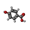  Mass: 138.121 Da / Num. of mol.: 1 / Source method: obtained synthetically / Formula: C7H6O3 Mass: 138.121 Da / Num. of mol.: 1 / Source method: obtained synthetically / Formula: C7H6O3 |
| #4: Water | ChemComp-HOH /  Mass: 18.015 Da / Num. of mol.: 218 / Source method: isolated from a natural source / Formula: H2O Mass: 18.015 Da / Num. of mol.: 218 / Source method: isolated from a natural source / Formula: H2O |
-Experimental details
-Experiment
| Experiment | Method: X-RAY DIFFRACTION / Number of used crystals: 1 |
|---|
- Sample preparation
| Crystal | Density Matthews: 2.7 Å3/Da / Density % sol: 53.72 % | |||||||||||||||||||||||||||||||||||||||||||||
|---|---|---|---|---|---|---|---|---|---|---|---|---|---|---|---|---|---|---|---|---|---|---|---|---|---|---|---|---|---|---|---|---|---|---|---|---|---|---|---|---|---|---|---|---|---|---|
| Crystal grow | pH: 7 / Details: pH 7.0 | |||||||||||||||||||||||||||||||||||||||||||||
| Components of the solutions |
| |||||||||||||||||||||||||||||||||||||||||||||
| Crystal grow | *PLUS Temperature: 4 ℃ / Method: vapor diffusion, hanging drop / Details: Eppink, M.H.M., (1995) Eur. J. Biochem., 231, 157. | |||||||||||||||||||||||||||||||||||||||||||||
| Components of the solutions | *PLUS
|
-Data collection
| Diffraction | Mean temperature: 298 K |
|---|---|
| Diffraction source | Source: ROTATING ANODE / Type: SIEMENS / Wavelength: 1.5418 |
| Detector | Type: SIEMENS / Detector: AREA DETECTOR / Date: Aug 15, 1996 / Details: COLLIMATOR |
| Radiation | Monochromator: GRAPHITE / Protocol: SINGLE WAVELENGTH / Monochromatic (M) / Laue (L): M / Scattering type: x-ray |
| Radiation wavelength | Wavelength: 1.5418 Å / Relative weight: 1 |
| Reflection | Resolution: 2.2→8 Å / Num. all: 22860 / Num. obs: 22860 / % possible obs: 92.5 % / Observed criterion σ(I): 0 / Redundancy: 2.5 % / Rsym value: 5.5 / Net I/σ(I): 12 |
| Reflection | *PLUS Rmerge(I) obs: 0.055 |
- Processing
| Software |
| ||||||||||||||||||||||||||||||||||||||||||||||||||||||||||||
|---|---|---|---|---|---|---|---|---|---|---|---|---|---|---|---|---|---|---|---|---|---|---|---|---|---|---|---|---|---|---|---|---|---|---|---|---|---|---|---|---|---|---|---|---|---|---|---|---|---|---|---|---|---|---|---|---|---|---|---|---|---|
| Refinement | Method to determine structure: MOLECULAR REPLACEMENT Starting model: 1PBE Resolution: 2.2→8 Å / Data cutoff high absF: 1000000 / Data cutoff low absF: 0.001 / Cross valid method: THROUGHOUT / σ(F): 0
| ||||||||||||||||||||||||||||||||||||||||||||||||||||||||||||
| Displacement parameters | Biso mean: 30 Å2 | ||||||||||||||||||||||||||||||||||||||||||||||||||||||||||||
| Refine analyze | Luzzati coordinate error obs: 0.28 Å | ||||||||||||||||||||||||||||||||||||||||||||||||||||||||||||
| Refinement step | Cycle: LAST / Resolution: 2.2→8 Å
| ||||||||||||||||||||||||||||||||||||||||||||||||||||||||||||
| Refine LS restraints |
| ||||||||||||||||||||||||||||||||||||||||||||||||||||||||||||
| Xplor file |
| ||||||||||||||||||||||||||||||||||||||||||||||||||||||||||||
| Software | *PLUS Name: X-PLOR / Version: 3.851 / Classification: refinement | ||||||||||||||||||||||||||||||||||||||||||||||||||||||||||||
| Refinement | *PLUS Highest resolution: 2.2 Å / Lowest resolution: 8 Å / σ(F): 0 / Rfactor obs: 0.185 | ||||||||||||||||||||||||||||||||||||||||||||||||||||||||||||
| Solvent computation | *PLUS | ||||||||||||||||||||||||||||||||||||||||||||||||||||||||||||
| Displacement parameters | *PLUS Biso mean: 30 Å2 | ||||||||||||||||||||||||||||||||||||||||||||||||||||||||||||
| Refine LS restraints | *PLUS
|
