THERE ARE CONFLICTS BETWEEN THE REPORTED SEQUENCE AND DATABASE REFERENCE ONE(P96558 IN UNIPROT). ...THERE ARE CONFLICTS BETWEEN THE REPORTED SEQUENCE AND DATABASE REFERENCE ONE(P96558 IN UNIPROT). ACCORDING TO THE DEPOSITORS, THE DIFFERENCE IN THE RESIDUES IS POSSIBLY DUE TO DIFFERENT ANNOTATION IN THE DATA BASE OR DUE TO THE CONSTRUCTION OF THE CHIMERIC PROTEIN. THIS COORDINATES USE NON-SEQUENTIAL RESIDUE NUMBERING FOR CLARIFICATION OF THE EXPRESSION TAGS AT THE C-TERMINUS.
-
実験情報
-
実験
実験
手法: X線回折 / 使用した結晶の数: 1
-
試料調製
結晶
マシュー密度: 2.87 Å3/Da / 溶媒含有率: 57.14 %
結晶化
温度: 300 K / 手法: 蒸気拡散法, ハンギングドロップ法 / pH: 8.5 詳細: Tris HCl pH 8.5, 1.2-2M ammonium sulphate, VAPOR DIFFUSION, HANGING DROP, temperature 300K
 ムービー
ムービー コントローラー


 データを開く
 基本情報
基本情報 要素
要素 キーワード
キーワード 機能・相同性情報
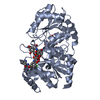
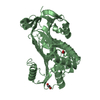
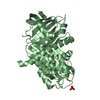
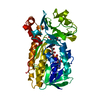
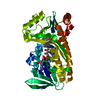
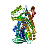
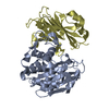
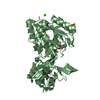
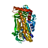
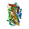
 Amycolatopsis orientalis (バクテリア)
 X線回折 /
  データ登録者
 引用
引用 構造の表示
構造の表示 ダウンロードとリンク
 その他のダウンロード
 PDBj
PDBj


 集合体
集合体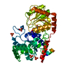

 分子量: 94.971 Da / 分子数: 4 / 由来タイプ: 合成 / 式: PO4
 タイプ: RNA linking / 分子量: 404.161 Da / 分子数: 1 / 由来タイプ: 合成 / 式: C9H14N2O12P2 / コメント: UDP*YM
 分子量: 18.015 Da / 分子数: 589 / 由来タイプ: 天然 / 式: H2O
 試料調製
試料調製 / ビームライン: BM14 / 波長: 0.977 Å
 解析
解析