 Movie
Movie Controller

[English] 日本語
 Yorodumi
- PDB-1o6z: 1.95 A resolution structure of (R207S,R292S) mutant of malate deh... -
+ Open data

- Basic information
| Entry | Database: PDB / ID: 1o6z | ||||||
|---|---|---|---|---|---|---|---|
| Title | 1.95 A resolution structure of (R207S,R292S) mutant of malate dehydrogenase from the halophilic archaeon Haloarcula marismortui (holo form) | ||||||
 Components Components | MALATE DEHYDROGENASE | ||||||
 Keywords Keywords | OXIDOREDUCTASE / HALOPHILIC / ION-BINDING / PROTEIN-SOLVENT INTERACTION / MALATE DEHYDROGENASE | ||||||
| Function / homology |  Function and homology information Function and homology information(S)-malate dehydrogenase (NAD+, oxaloacetate-forming) / L-malate dehydrogenase (NAD+) activity / L-lactate dehydrogenase (NAD+) activity / lactate metabolic process / tricarboxylic acid cycle / cytoplasm Similarity search - Function | ||||||
| Biological species |  HALOARCULA MARISMORTUI (Halophile) HALOARCULA MARISMORTUI (Halophile) | ||||||
| Method |  X-RAY DIFFRACTION / SYNCHROTRON / MOLECULAR REPLACEMENT / Resolution: 1.95 Å X-RAY DIFFRACTION / SYNCHROTRON / MOLECULAR REPLACEMENT / Resolution: 1.95 Å | ||||||
 Authors Authors | Irimia, A. / Ebel, C. / Madern, D. / Richard, S.B. / Cosenza, L.W. / Zaccai, G. / Vellieux, F.M.D. | ||||||
 Citation Citation | Journal: J.Mol.Biol. / Year: 2003 Title: The Oligomeric States of Haloarcula Marismortui Malate Dehydrogenase are Modulated by Solvent Components as Shown by Crystallographic and Biochemical Studies Authors: Irimia, A. / Ebel, C. / Madern, D. / Richard, S.B. / Cosenza, L.W. / Zaccai, G. / Vellieux, F.M.D. #1: Journal: Biochemistry / Year: 2000 Title: Insights Into the Molecular Relationships between Malate and Lactate Dehydrogenases: Structural and Biochemical Properties of Monomeric and Dimeric Intermediates of a Mutant of Tetrameric L- ...Title: Insights Into the Molecular Relationships between Malate and Lactate Dehydrogenases: Structural and Biochemical Properties of Monomeric and Dimeric Intermediates of a Mutant of Tetrameric L-[Ldh-Like] Malate Dehydrogenase from the Halophilic Archaeon Haloarcula Marismortui Authors: Madern, D. / Ebel, C. / Mevarech, M. / Richard, S.B. / Pfister, C. / Zaccai, G. #2: Journal: Biochemistry / Year: 2000Title: Halophilic Adaptation: Novel Solvent Protein Interactions Observed in the 2.9 And 2.6 A Resolution Structures of the Wild Type and a Mutant of Malate Dehydrogenase from Haloarcula Marismortui Authors: Richard, S.B. / Madern, D. / Garcin, E. / Zaccai, G. #3: Journal: Science / Year: 1995Title: Structural Features that Stabilize Halophilic Malate Dehydrogenase from an Archaebacterium Authors: Dym, O. / Mevarech, M. / Sussman, J.L. #4: Journal: Eur.J.Biochem. / Year: 1995 Title: Mutation at a Single Acidic Amino Acid Enhances the Halophilic Behaviour of Malate Dehydrogenase from Haloarcula Marismortui in Physiological Salts Authors: Madern, D. / Pfister, C. / Zaccai, G. #5: Journal: Biochemistry / Year: 1993 Title: Cloning, Sequencing, and Expression in Escherichia Coli of the Gene Coding for Malate Dehydrogenase of the Extremely Halophilic Archaebacterium Haloarcula Marismortui Authors: Cendrin, F. / Chroboczek, J. / Zaccai, G. / Eisenberg, H. / Mevarech, M. | ||||||
| History |
|
- Structure visualization
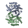
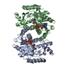
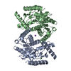
| Structure viewer | Molecule: MolmilJmol/JSmol |
|---|
- Downloads & links
-Download
| PDBx/mmCIF format | 1o6z.cif.gz | 254.5 KB | Display | PDBx/mmCIF format |
|---|---|---|---|---|
| PDB format | pdb1o6z.ent.gz | 205.3 KB | Display | PDB format |
| PDBx/mmJSON format | 1o6z.json.gz | Tree view | PDBx/mmJSON format | |
| Others |  Other downloads Other downloads |
-Validation report
| Arichive directory | https://data.pdbj.org/pub/pdb/validation_reports/o6/1o6zftp://data.pdbj.org/pub/pdb/validation_reports/o6/1o6z | HTTPS FTP |
|---|
-Related structure data
| Related structure data |  2x0rC  1gt2 C: citing same article ( S: Starting model for refinement |
|---|---|
| Similar structure data |


-Links
 PDBj
PDBj


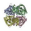
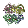
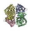
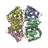
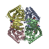
- Assembly
| Deposited unit | 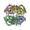
| ||||||||||||||||||||||||||||
|---|---|---|---|---|---|---|---|---|---|---|---|---|---|---|---|---|---|---|---|---|---|---|---|---|---|---|---|---|---|
| 1 |
| ||||||||||||||||||||||||||||
| Unit cell |
| ||||||||||||||||||||||||||||
| Components on special symmetry positions |
| ||||||||||||||||||||||||||||
| Noncrystallographic symmetry (NCS) | NCS oper:
|
-Components
| #1: Protein | Mass: 32566.301 Da / Num. of mol.: 4 / Mutation: YES Source method: isolated from a genetically manipulated source Details: COMPLEXED WITH NADH / Source: (gene. exp.) HALOARCULA MARISMORTUI (Halophile) / Plasmid: PET11A / Production host:  References: UniProt: Q07841, (S)-malate dehydrogenase (NAD+, oxaloacetate-forming) #2: Chemical | ChemComp-CL /   Mass: 35.453 Da / Num. of mol.: 8 / Source method: obtained synthetically / Formula: Cl Mass: 35.453 Da / Num. of mol.: 8 / Source method: obtained synthetically / Formula: Cl#3: Chemical | ChemComp-NAD /   Mass: 663.425 Da / Num. of mol.: 4 / Source method: obtained synthetically / Formula: C21H27N7O14P2 / Comment: NAD*YM Mass: 663.425 Da / Num. of mol.: 4 / Source method: obtained synthetically / Formula: C21H27N7O14P2 / Comment: NAD*YM#4: Water | ChemComp-HOH / |  Mass: 18.015 Da / Num. of mol.: 677 / Source method: isolated from a natural source / Formula: H2O Mass: 18.015 Da / Num. of mol.: 677 / Source method: isolated from a natural source / Formula: H2OCompound details | ENGINEERED | |
|---|
-Experimental details
-Experiment
| Experiment | Method: X-RAY DIFFRACTION / Number of used crystals: 1 |
|---|
- Sample preparation
| Crystal | Density Matthews: 3.46 Å3/Da / Density % sol: 62.41 % | |||||||||||||||||||||||||||||||||||
|---|---|---|---|---|---|---|---|---|---|---|---|---|---|---|---|---|---|---|---|---|---|---|---|---|---|---|---|---|---|---|---|---|---|---|---|---|
| Crystal grow | pH: 7.6 / Details: 2 M NACL, 25 MM TRIS PH 7.6, 2.5 MM NADH, 50% MPD | |||||||||||||||||||||||||||||||||||
| Crystal grow | *PLUS Temperature: 4 ℃ / Method: vapor diffusion, sitting drop | |||||||||||||||||||||||||||||||||||
| Components of the solutions | *PLUS
|
-Data collection
| Diffraction | Mean temperature: 100 K |
|---|---|
| Diffraction source | Source: SYNCHROTRON / Site: ESRF  / Beamline: ID14-2 / Wavelength: 0.933 / Beamline: ID14-2 / Wavelength: 0.933 |
| Detector | Type: ADSC CCD / Detector: CCD / Date: Jun 17, 2000 / Details: TOROIDAL MIRROR |
| Radiation | Monochromator: GE(220) / Protocol: SINGLE WAVELENGTH / Monochromatic (M) / Laue (L): M / Scattering type: x-ray |
| Radiation wavelength | Wavelength: 0.933 Å / Relative weight: 1 |
| Reflection | Resolution: 1.95→42.26 Å / Num. obs: 100475 / % possible obs: 77.8 % / Observed criterion σ(I): 0 / Redundancy: 3.34 % / Biso Wilson estimate: 18.84 Å2 / Rmerge(I) obs: 0.0642 / Net I/σ(I): 12.86 |
| Reflection shell | Resolution: 1.95→2.02 Å / Redundancy: 2.12 % / Rmerge(I) obs: 0.1427 / Mean I/σ(I) obs: 4.03 / % possible all: 43.9 |
| Reflection | *PLUS Num. measured all: 335871 |
| Reflection shell | *PLUS % possible obs: 43.9 % / Num. unique obs: 5703 / Num. measured obs: 12111 |
- Processing
| Software |
| ||||||||||||||||||||
|---|---|---|---|---|---|---|---|---|---|---|---|---|---|---|---|---|---|---|---|---|---|
| Refinement | Method to determine structure: MOLECULAR REPLACEMENT Starting model: PDB ENTRY 1GT2 1gt2 Resolution: 1.95→12 Å / SU B: 6.089 / SU ML: 0.167 / Cross valid method: THROUGHOUT / ESU R: 0.26 / ESU R Free: 0.17 Details: HYDROGENS HAVE BEEN ADDED IN THE RIDING POSITIONS. THE REGION 100-107 OF B AND D CHAIN IS DISORDERED. THE RESIDUES 101-106 IN THESE DISORDERED REGIONS COULD NOT BE MODELLED AND ARE OMITTED FROM THE MODEL.
| ||||||||||||||||||||
| Displacement parameters | Biso mean: 24.478 Å2
| ||||||||||||||||||||
| Refinement step | Cycle: LAST / Resolution: 1.95→12 Å
| ||||||||||||||||||||
| Refinement | *PLUS Lowest resolution: 42.26 Å / Num. reflection obs: 95398 / Num. reflection Rfree: 5076 / Rfactor Rfree: 0.263 / Rfactor Rwork: 0.19 | ||||||||||||||||||||
| Solvent computation | *PLUS | ||||||||||||||||||||
| Displacement parameters | *PLUS | ||||||||||||||||||||
| Refine LS restraints | *PLUS
| ||||||||||||||||||||
| LS refinement shell | *PLUS Highest resolution: 1.955 Å / Lowest resolution: 2.005 Å / Rfactor Rfree: 0.311 / % reflection Rfree: 204 % / Rfactor Rwork: 0.213 / Num. reflection Rwork: 4065 / Total num. of bins used: 20 |
