 Movie
Movie Controller

[English] 日本語
 Yorodumi
- PDB-1d3a: CRYSTAL STRUCTURE OF THE WILD TYPE HALOPHILIC MALATE DEHYDROGENAS... -
+ Open data

- Basic information
| Entry | Database: PDB / ID: 1d3a | ||||||
|---|---|---|---|---|---|---|---|
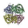
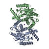
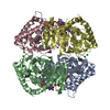
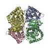
| Title | CRYSTAL STRUCTURE OF THE WILD TYPE HALOPHILIC MALATE DEHYDROGENASE IN THE APO FORM | ||||||
 Components Components | HALOPHILIC MALATE DEHYDROGENASE | ||||||
 Keywords Keywords | OXIDOREDUCTASE / ROSSMANN FOLD AND 3 SORTS OF COMPLEX SALT BRIDGES | ||||||
| Function / homology |  Function and homology information Function and homology information(S)-malate dehydrogenase (NAD+, oxaloacetate-forming) / L-malate dehydrogenase (NAD+) activity / L-lactate dehydrogenase (NAD+) activity / lactate metabolic process / tricarboxylic acid cycle / cytoplasm Similarity search - Function | ||||||
| Biological species |  Haloarcula marismortui (Halophile) Haloarcula marismortui (Halophile) | ||||||
| Method |  X-RAY DIFFRACTION / SYNCHROTRON / Resolution: 2.94 Å X-RAY DIFFRACTION / SYNCHROTRON / Resolution: 2.94 Å | ||||||
 Authors Authors | Richard, S.B. / Madern, D. / Garcin, E. / Zaccai, G. | ||||||
 Citation Citation | Journal: Biochemistry / Year: 2000 Title: Halophilic adaptation: novel solvent protein interactions observed in the 2.9 and 2.6 A resolution structures of the wild type and a mutant of malate dehydrogenase from Haloarcula marismortui. Authors: Richard, S.B. / Madern, D. / Garcin, E. / Zaccai, G. #1: Journal: ARCHAEA : A LABORATORY MANUAL. V.[1]. HALOPHILES / Year: 1995Title: Protocol 21: the MPD-NACL-H2O System for the Crystallization of Halophilic Proteins Authors: Richard, S.B. / Bonnete, F. / Dym, O. / Zaccai, G. #2: Journal: Science / Year: 1995Title: Structural Features Stabilizing Halophilic Malate Dehydrogenase from an Archaebacterium Authors: Dym, O. / Mevarech, M. / Sussman, J.L. #3: Journal: Eur.J.Biochem. / Year: 1995Title: Mutation at a Single Amino Acid Enhances the Halophilic Behaviour of Malate Dehydrogenase from Haloarcula Marismortui in physiological salts Authors: Madern, D. / Pfister, C. / Zaccai, G. #4: Journal: Biochemistry / Year: 1993Title: Cloning, Sequencing, and Expression in Escherichia Coli of the Gene Coding for Malate Dehydrogenase of the Extremely Halophilic Archaebacterium Haloarcula Marismortui Authors: Cendrin, F. / Chroboczek, J. / Zaccai, G. / Eisenberg, H. / Mevarech, M. | ||||||
| History |
|
- Structure visualization
| Structure viewer | Molecule: MolmilJmol/JSmol |
|---|
- Downloads & links
-Download
| PDBx/mmCIF format | 1d3a.cif.gz | 124.1 KB | Display | PDBx/mmCIF format |
|---|---|---|---|---|
| PDB format | pdb1d3a.ent.gz | 97.6 KB | Display | PDB format |
| PDBx/mmJSON format | 1d3a.json.gz | Tree view | PDBx/mmJSON format | |
| Others |  Other downloads Other downloads |
-Validation report
| Arichive directory | https://data.pdbj.org/pub/pdb/validation_reports/d3/1d3aftp://data.pdbj.org/pub/pdb/validation_reports/d3/1d3a | HTTPS FTP |
|---|
-Related structure data


-Links
 PDBj
PDBj


- Assembly
| Deposited unit | 
| ||||||||
|---|---|---|---|---|---|---|---|---|---|
| 1 | 
| ||||||||
| Unit cell |
|
-Components
| #1: Protein | Mass: 32706.531 Da / Num. of mol.: 2 Source method: isolated from a genetically manipulated source Source: (gene. exp.) Haloarcula marismortui (Halophile) / Plasmid: PET11D / Production host:  References: UniProt: Q07841, (S)-malate dehydrogenase (NAD+, oxaloacetate-forming) #2: Chemical |   Mass: 35.453 Da / Num. of mol.: 2 / Source method: obtained synthetically / Formula: Cl Mass: 35.453 Da / Num. of mol.: 2 / Source method: obtained synthetically / Formula: Cl#3: Chemical | ChemComp-NA / |   Mass: 22.990 Da / Num. of mol.: 1 / Source method: obtained synthetically / Formula: Na Mass: 22.990 Da / Num. of mol.: 1 / Source method: obtained synthetically / Formula: Na#4: Water | ChemComp-HOH / |  Mass: 18.015 Da / Num. of mol.: 32 / Source method: isolated from a natural source / Formula: H2O Mass: 18.015 Da / Num. of mol.: 32 / Source method: isolated from a natural source / Formula: H2O |
|---|
-Experimental details
-Experiment
| Experiment | Method: X-RAY DIFFRACTION / Number of used crystals: 3 |
|---|
- Sample preparation
| Crystal | Density Matthews: 3.64 Å3/Da / Density % sol: 66.24 % | ||||||||||||||||||||||||||||||||||||||||||
|---|---|---|---|---|---|---|---|---|---|---|---|---|---|---|---|---|---|---|---|---|---|---|---|---|---|---|---|---|---|---|---|---|---|---|---|---|---|---|---|---|---|---|---|
| Crystal grow | Temperature: 298 K / Method: vapor diffusion, sitting drop / pH: 7.6 Details: MPD-NaCl-H2O system, pH 7.6, VAPOR DIFFUSION, SITTING DROP, temperature 298K | ||||||||||||||||||||||||||||||||||||||||||
| Crystal | *PLUS Density % sol: 70 % | ||||||||||||||||||||||||||||||||||||||||||
| Crystal grow | *PLUS Temperature: 6 ℃ | ||||||||||||||||||||||||||||||||||||||||||
| Components of the solutions | *PLUS
|
-Data collection
| Diffraction | Mean temperature: 284 K |
|---|---|
| Diffraction source | Source: SYNCHROTRON / Site: LURE  / Beamline: DW32 / Wavelength: 0.975 / Beamline: DW32 / Wavelength: 0.975 |
| Detector | Type: MARRESEARCH / Detector: IMAGE PLATE / Date: Oct 22, 1998 |
| Radiation | Protocol: SINGLE WAVELENGTH / Monochromatic (M) / Laue (L): M / Scattering type: x-ray |
| Radiation wavelength | Wavelength: 0.975 Å / Relative weight: 1 |
| Reflection | Resolution: 2.94→30 Å / Num. obs: 99423 / % possible obs: 94.2 % / Observed criterion σ(I): 2 / Redundancy: 4.9 % / Biso Wilson estimate: 82.9 Å2 / Rmerge(I) obs: 0.1 / Net I/σ(I): 3.2 |
| Reflection shell | Resolution: 2.94→3.07 Å / Redundancy: 5.2 % / Rmerge(I) obs: 0.332 / Num. unique all: 19960 / % possible all: 94.9 |
| Reflection | *PLUS Num. obs: 19960 / Num. measured all: 99423 |
| Reflection shell | *PLUS % possible obs: 94.5 % / Num. unique obs: 3237 |
- Processing
| Software |
| ||||||||||||||||||||||||||||||||||||
|---|---|---|---|---|---|---|---|---|---|---|---|---|---|---|---|---|---|---|---|---|---|---|---|---|---|---|---|---|---|---|---|---|---|---|---|---|---|
| Refinement | Resolution: 2.94→30 Å / Rfactor Rfree error: 0.016 / Data cutoff high absF: 2600643.31 / Data cutoff low absF: 0 / Isotropic thermal model: RESTRAINED / Cross valid method: THROUGHOUT / σ(F): 0 / Stereochemistry target values: Engh & Huber / Details: using maximum likelihood target using amplitudes
| ||||||||||||||||||||||||||||||||||||
| Solvent computation | Solvent model: FLAT MODEL | ||||||||||||||||||||||||||||||||||||
| Displacement parameters | Biso mean: 32.2 Å2
| ||||||||||||||||||||||||||||||||||||
| Refine analyze |
| ||||||||||||||||||||||||||||||||||||
| Refinement step | Cycle: LAST / Resolution: 2.94→30 Å
| ||||||||||||||||||||||||||||||||||||
| Refine LS restraints |
| ||||||||||||||||||||||||||||||||||||
| LS refinement shell | Resolution: 2.94→3.04 Å / Rfactor Rfree error: 0.036 / Total num. of bins used: 6
| ||||||||||||||||||||||||||||||||||||
| Xplor file |
| ||||||||||||||||||||||||||||||||||||
| Software | *PLUS Name: CNS / Version: 0.4 / Classification: refinement | ||||||||||||||||||||||||||||||||||||
| Refinement | *PLUS Rfactor Rwork: 0.188 / Num. reflection obs: 15920 | ||||||||||||||||||||||||||||||||||||
| Solvent computation | *PLUS | ||||||||||||||||||||||||||||||||||||
| Displacement parameters | *PLUS Biso mean: 61.5 Å2 | ||||||||||||||||||||||||||||||||||||
| Refine LS restraints | *PLUS
|
