 Movie
Movie Controller

[English] 日本語
 Yorodumi
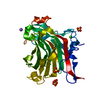
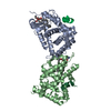
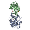
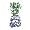
- PDB-1o4z: THE THREE-DIMENSIONAL STRUCTURE OF BETA-AGARASE B FROM ZOBELLIA G... -
+ Open data

- Basic information
| Entry | Database: PDB / ID: 1o4z | ||||||
|---|---|---|---|---|---|---|---|
| Title | THE THREE-DIMENSIONAL STRUCTURE OF BETA-AGARASE B FROM ZOBELLIA GALACTANIVORANS | ||||||
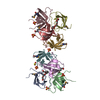
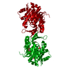
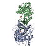
 Components Components | beta-agarase B | ||||||
 Keywords Keywords | HYDROLASE / BETA-AGARASE / GLYCOSIDE HYDROLASE FAMILY 16 / AGAROSE DEGRADATION / CLEAVAGE OF BETA-1 / 4-D-GALACTOSE LINKAGES | ||||||
| Function / homology |  Function and homology information Function and homology informationbeta-agarase / beta-agarase activity / cell outer membrane / carbohydrate metabolic process / protein homodimerization activity Similarity search - Function | ||||||
| Biological species |  Zobellia galactanivorans (bacteria) Zobellia galactanivorans (bacteria) | ||||||
| Method |  X-RAY DIFFRACTION / SYNCHROTRON / MOLECULAR REPLACEMENT / Resolution: 2.3 Å X-RAY DIFFRACTION / SYNCHROTRON / MOLECULAR REPLACEMENT / Resolution: 2.3 Å | ||||||
 Authors Authors | Allouch, J. / Jam, M. / Helbert, W. / Barbeyron, T. / Kloareg, B. / Henrissat, B. / Czjzek, M. | ||||||
 Citation Citation | Journal: J.Biol.Chem. / Year: 2003 Title: The Three-dimensional Structures of Two {beta}-Agarases. Authors: Allouch, J. / Jam, M. / Helbert, W. / Barbeyron, T. / Kloareg, B. / Henrissat, B. / Czjzek, M. | ||||||
| History |
|
- Structure visualization
| Structure viewer | Molecule: MolmilJmol/JSmol |
|---|
- Downloads & links
-Download
| PDBx/mmCIF format | 1o4z.cif.gz | 272.5 KB | Display | PDBx/mmCIF format |
|---|---|---|---|---|
| PDB format | pdb1o4z.ent.gz | 216 KB | Display | PDB format |
| PDBx/mmJSON format | 1o4z.json.gz | Tree view | PDBx/mmJSON format | |
| Others |  Other downloads Other downloads |
-Validation report
| Summary document | 1o4z_validation.pdf.gz | 483 KB | Display | wwPDB validaton report |
|---|---|---|---|---|
| Full document | 1o4z_full_validation.pdf.gz | 505.4 KB | Display | |
| Data in XML | 1o4z_validation.xml.gz | 55.5 KB | Display | |
| Data in CIF | 1o4z_validation.cif.gz | 79.4 KB | Display | |
| Arichive directory | https://data.pdbj.org/pub/pdb/validation_reports/o4/1o4zftp://data.pdbj.org/pub/pdb/validation_reports/o4/1o4z | HTTPS FTP |
-Related structure data


-Links
 PDBj
PDBj


- Assembly
| Deposited unit | 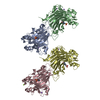
| ||||||||||||||||
|---|---|---|---|---|---|---|---|---|---|---|---|---|---|---|---|---|---|
| 1 | 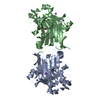
| ||||||||||||||||
| 2 | 
| ||||||||||||||||
| Unit cell |
| ||||||||||||||||
| Noncrystallographic symmetry (NCS) | NCS oper:
|
-Components
| #1: Protein | Mass: 40056.523 Da / Num. of mol.: 4 Source method: isolated from a genetically manipulated source Source: (gene. exp.) Zobellia galactanivorans (bacteria) / Strain: DSIJ / Gene: agaB / Plasmid: PET20B / Production host: #2: Chemical | ChemComp-NA /   Mass: 22.990 Da / Num. of mol.: 7 / Source method: obtained synthetically / Formula: Na Mass: 22.990 Da / Num. of mol.: 7 / Source method: obtained synthetically / Formula: Na#3: Chemical | ChemComp-EPE /   Mass: 238.305 Da / Num. of mol.: 4 / Source method: obtained synthetically / Formula: C8H18N2O4S / Comment: pH buffer*YM Mass: 238.305 Da / Num. of mol.: 4 / Source method: obtained synthetically / Formula: C8H18N2O4S / Comment: pH buffer*YM#4: Chemical |   Mass: 24.305 Da / Num. of mol.: 2 / Source method: obtained synthetically / Formula: Mg Mass: 24.305 Da / Num. of mol.: 2 / Source method: obtained synthetically / Formula: Mg#5: Water | ChemComp-HOH / |  Mass: 18.015 Da / Num. of mol.: 888 / Source method: isolated from a natural source / Formula: H2O Mass: 18.015 Da / Num. of mol.: 888 / Source method: isolated from a natural source / Formula: H2O |
|---|
-Experimental details
-Experiment
| Experiment | Method: X-RAY DIFFRACTION / Number of used crystals: 1 |
|---|
- Sample preparation
| Crystal | Density Matthews: 2.31 Å3/Da / Density % sol: 47 % | |||||||||||||||||||||||||||||||||||||||||||||||||
|---|---|---|---|---|---|---|---|---|---|---|---|---|---|---|---|---|---|---|---|---|---|---|---|---|---|---|---|---|---|---|---|---|---|---|---|---|---|---|---|---|---|---|---|---|---|---|---|---|---|---|
| Crystal grow | Method: vapor diffusion, hanging drop / pH: 7.5 Details: 58% methyl-pentane-diol 20 mM calcium chloride 100 mM Hepes , pH 7.5, VAPOR DIFFUSION, HANGING DROP | |||||||||||||||||||||||||||||||||||||||||||||||||
| Crystal grow | *PLUS Temperature: 20.5 ℃ / Method: vapor diffusion, hanging drop | |||||||||||||||||||||||||||||||||||||||||||||||||
| Components of the solutions | *PLUS
|
-Data collection
| Diffraction | Mean temperature: 100 K |
|---|---|
| Diffraction source | Source: SYNCHROTRON / Site: ESRF  / Beamline: ID14-2 / Wavelength: 0.933 / Beamline: ID14-2 / Wavelength: 0.933 |
| Detector | Type: ADSC QUANTUM 4 / Detector: CCD / Date: Dec 1, 2002 |
| Radiation | Protocol: SINGLE WAVELENGTH / Monochromatic (M) / Laue (L): M / Scattering type: x-ray |
| Radiation wavelength | Wavelength: 0.933 Å / Relative weight: 1 |
| Reflection | Resolution: 2.3→95.35 Å / Num. all: 61473 / Num. obs: 61473 / % possible obs: 99.9 % / Observed criterion σ(I): 0 / Redundancy: 3.5 % / Rsym value: 0.106 / Net I/σ(I): 5.4 |
| Reflection shell | Resolution: 2.3→2.42 Å / Redundancy: 3.4 % / Mean I/σ(I) obs: 1.6 / Rsym value: 0.339 / % possible all: 99.8 |
| Reflection | *PLUS Lowest resolution: 24.8 Å / Num. obs: 64783 / Num. measured all: 428617 / Rmerge(I) obs: 0.106 |
| Reflection shell | *PLUS % possible obs: 99.9 % / Rmerge(I) obs: 0.339 |
- Processing
| Software |
| |||||||||||||||||||||||||||||||||||||||||||||||||||||||||||||||
|---|---|---|---|---|---|---|---|---|---|---|---|---|---|---|---|---|---|---|---|---|---|---|---|---|---|---|---|---|---|---|---|---|---|---|---|---|---|---|---|---|---|---|---|---|---|---|---|---|---|---|---|---|---|---|---|---|---|---|---|---|---|---|---|---|
| Refinement | Method to determine structure: MOLECULAR REPLACEMENT Starting model: BETA-AGARASE A Resolution: 2.3→25 Å / SU B: 5.974 / SU ML: 0.145 / Cross valid method: THROUGHOUT / σ(F): 0 / ESU R: 0.292 / ESU R Free: 0.218
| |||||||||||||||||||||||||||||||||||||||||||||||||||||||||||||||
| Displacement parameters | Biso mean: 19.944 Å2
| |||||||||||||||||||||||||||||||||||||||||||||||||||||||||||||||
| Refinement step | Cycle: LAST / Resolution: 2.3→25 Å
| |||||||||||||||||||||||||||||||||||||||||||||||||||||||||||||||
| Refine LS restraints |
| |||||||||||||||||||||||||||||||||||||||||||||||||||||||||||||||
| Refinement | *PLUS Lowest resolution: 24.8 Å / Rfactor Rfree: 0.224 / Rfactor Rwork: 0.199 | |||||||||||||||||||||||||||||||||||||||||||||||||||||||||||||||
| Solvent computation | *PLUS | |||||||||||||||||||||||||||||||||||||||||||||||||||||||||||||||
| Displacement parameters | *PLUS | |||||||||||||||||||||||||||||||||||||||||||||||||||||||||||||||
| Refine LS restraints | *PLUS
|
