% possible obs: 95.3 % / Rmerge(I) obs: 0.196 / Mean I/σ(I) obs: 3.5
-
Processing
Software
Name
Version
Classification
DENZO
datareduction
SCALEPACK
datascaling
RESOLVE
modelbuilding
SOLVE
phasing
ARP/wARP
modelbuilding
REFMAC
5.1.955
refinement
RESOLVE
phasing
Refinement
Method to determine structure: MAD / Resolution: 1.6→48.18 Å / Cor.coef. Fo:Fc: 0.974 / Cor.coef. Fo:Fc free: 0.956 / SU B: 2.507 / SU ML: 0.041 / TLS residual ADP flag: LIKELY RESIDUAL / Isotropic thermal model: Isotropic / Cross valid method: THROUGHOUT / σ(F): 0 / ESU R: 0.067 / ESU R Free: 0.075 / Stereochemistry target values: MAXIMUM LIKELIHOOD Details: WEAK CONTINUOUS DIFFERENCE DENSITY AT THE N-TERMINUS WAS MODELLED AS HIS-TAG RESIDUES, BUT AT HALF OCCUPANCY.THE CONTINUED PRESENCE OF SPURIOUS DIFFERENCE DENSITY IMPLIES THAT THIS STILL IS ...Details: WEAK CONTINUOUS DIFFERENCE DENSITY AT THE N-TERMINUS WAS MODELLED AS HIS-TAG RESIDUES, BUT AT HALF OCCUPANCY.THE CONTINUED PRESENCE OF SPURIOUS DIFFERENCE DENSITY IMPLIES THAT THIS STILL IS AN INCOMPLETE DESCRIPTION OF THE N-TERMINUS. XFIT/CCP4/TLS WAS ALSO USED IN REFINEMENT.
Rfactor
Num. reflection
% reflection
Selection details
Rfree
0.18
1926
5.1 %
RANDOM
Rwork
0.139
-
-
-
obs
0.141
36027
97.8 %
-
Solvent computation
Ion probe radii: 0.8 Å / Shrinkage radii: 0.8 Å / VDW probe radii: 1.4 Å / Solvent model: BABINET MODEL WITH MASK
Displacement parameters
Biso mean: 13.32 Å2
Baniso -1
Baniso -2
Baniso -3
1-
0 Å2
0 Å2
0 Å2
2-
-
0.06 Å2
0 Å2
3-
-
-
-0.06 Å2
Refinement step
Cycle: LAST / Resolution: 1.6→48.18 Å
Protein
Nucleic acid
Ligand
Solvent
Total
Num. atoms
1831
0
1
422
2254
Refine LS restraints
Refine-ID
Type
Dev ideal
Dev ideal target
Number
X-RAY DIFFRACTION
r_bond_refined_d
0.012
0.021
1909
X-RAY DIFFRACTION
r_bond_other_d
0.002
0.02
1796
X-RAY DIFFRACTION
r_angle_refined_deg
1.411
1.969
2571
X-RAY DIFFRACTION
r_angle_other_deg
0.882
3
4177
X-RAY DIFFRACTION
r_dihedral_angle_1_deg
6.079
5
225
X-RAY DIFFRACTION
r_dihedral_angle_2_deg
34.05
24.124
97
X-RAY DIFFRACTION
r_dihedral_angle_3_deg
10.828
15
376
X-RAY DIFFRACTION
r_dihedral_angle_4_deg
19.046
15
15
X-RAY DIFFRACTION
r_chiral_restr
0.091
0.2
296
X-RAY DIFFRACTION
r_gen_planes_refined
0.006
0.02
2072
X-RAY DIFFRACTION
r_gen_planes_other
0.001
0.02
390
X-RAY DIFFRACTION
r_nbd_refined
0.211
0.2
383
X-RAY DIFFRACTION
r_nbd_other
0.238
0.2
2207
X-RAY DIFFRACTION
r_nbtor_refined
X-RAY DIFFRACTION
r_nbtor_other
0.08
0.2
1177
X-RAY DIFFRACTION
r_xyhbond_nbd_refined
0.23
0.2
248
X-RAY DIFFRACTION
r_xyhbond_nbd_other
X-RAY DIFFRACTION
r_metal_ion_refined
0.115
0.2
3
X-RAY DIFFRACTION
r_metal_ion_other
X-RAY DIFFRACTION
r_symmetry_vdw_refined
0.306
0.2
15
X-RAY DIFFRACTION
r_symmetry_vdw_other
0.336
0.2
48
X-RAY DIFFRACTION
r_symmetry_hbond_refined
0.244
0.2
45
X-RAY DIFFRACTION
r_symmetry_hbond_other
X-RAY DIFFRACTION
r_mcbond_it
1.79
4
1129
X-RAY DIFFRACTION
r_mcangle_it
3.054
6
1841
X-RAY DIFFRACTION
r_scbond_it
4.411
10
780
X-RAY DIFFRACTION
r_scangle_it
6.905
13
730
X-RAY DIFFRACTION
r_rigid_bond_restr
X-RAY DIFFRACTION
r_sphericity_free
X-RAY DIFFRACTION
r_sphericity_bonded
LS refinement shell
Resolution: 1.6→1.64 Å / Total num. of bins used: 20 /
Rfactor
Num. reflection
Rfree
0.218
137
Rwork
0.165
2413
Refinement TLS params.
Method: refined / Origin x: 20.0943 Å / Origin y: 8.3707 Å / Origin z: 2.9538 Å
In the structure databanks used in Yorodumi, some data are registered as the other names, "COVID-19 virus" and "2019-nCoV". Here are the details of the virus and the list of structure data.
Jan 31, 2019. EMDB accession codes are about to change! (news from PDBe EMDB page)
EMDB accession codes are about to change! (news from PDBe EMDB page)
The allocation of 4 digits for EMDB accession codes will soon come to an end. Whilst these codes will remain in use, new EMDB accession codes will include an additional digit and will expand incrementally as the available range of codes is exhausted. The current 4-digit format prefixed with “EMD-” (i.e. EMD-XXXX) will advance to a 5-digit format (i.e. EMD-XXXXX), and so on. It is currently estimated that the 4-digit codes will be depleted around Spring 2019, at which point the 5-digit format will come into force.
The EM Navigator/Yorodumi systems omit the EMD- prefix.
Related info.:Q: What is EMD? / ID/Accession-code notation in Yorodumi/EM Navigator
Yorodumi is a browser for structure data from EMDB, PDB, SASBDB, etc.
This page is also the successor to EM Navigator detail page, and also detail information page/front-end page for Omokage search.
The word "yorodu" (or yorozu) is an old Japanese word meaning "ten thousand". "mi" (miru) is to see.
Related info.:EMDB / PDB / SASBDB / Comparison of 3 databanks / Yorodumi Search / Aug 31, 2016. New EM Navigator & Yorodumi / Yorodumi Papers / Jmol/JSmol / Function and homology information / Changes in new EM Navigator and Yorodumi
 Movie
Movie Controller

 Yorodumi
 Open data
 Basic information
 Components
 Keywords
 Function and homology information
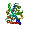
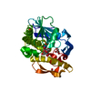
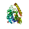
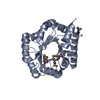
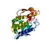
 Thermotoga maritima (bacteria)
 X-RAY DIFFRACTION /
  Authors
 Citation
 Structure visualization
 Downloads & links
 Other downloads
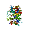


 PDBj
PDBj Assembly
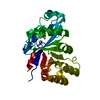

 Mass: 22.990 Da / Num. of mol.: 1 / Source method: obtained synthetically / Formula: Na
 Mass: 18.015 Da / Num. of mol.: 422 / Source method: isolated from a natural source / Formula: H2O
 Sample preparation
 / Beamline: 19-BM / Wavelength: 0.98011, 0.97938, 0.97918, 0.95671
 Processing