 Movie
Movie Controller

[English] 日本語
 Yorodumi
- PDB-2od6: Crystal structure of a dimeric ferredoxin-like protein (jcvi_pep_... -
+ Open data

- Basic information
| Entry | Database: PDB / ID: 2od6 | ||||||
|---|---|---|---|---|---|---|---|
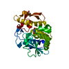
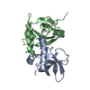
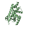
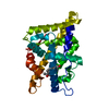
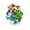
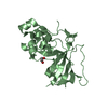
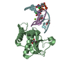
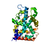
| Title | Crystal structure of a dimeric ferredoxin-like protein (jcvi_pep_1096682647733) from uncultured marine organism at 1.85 A resolution | ||||||
 Components Components | hypothetical protein | ||||||
 Keywords Keywords | UNKNOWN FUNCTION / Metagenomics target / structural genomics / Joint Center for Structural Genomics / JCSG / Protein Structure Initiative / PSI-2 | ||||||
| Function / homology | Alpha-Beta Plaits - #100 / Alpha-Beta Plaits / 2-Layer Sandwich / Alpha Beta / 10-oxohexadecanoic acid Function and homology information Function and homology information | ||||||
| Biological species | uncultured marine organism (environmental samples) | ||||||
| Method |  X-RAY DIFFRACTION / SYNCHROTRON / MAD / Resolution: 1.85 Å X-RAY DIFFRACTION / SYNCHROTRON / MAD / Resolution: 1.85 Å | ||||||
 Authors Authors | Joint Center for Structural Genomics (JCSG) | ||||||
 Citation Citation | Journal: To be published Title: Crystal structure of hypothetical protein (JCVI_PEP_1096682647733) from an environmental metagenome (unidentified marine microbe), Sorcerer II Global Ocean Sampling experiment at 1.85 A resolution Authors: Joint Center for Structural Genomics (JCSG) | ||||||
| History |
| ||||||
| Remark 300 | BIOMOLECULE: 1,2 THIS ENTRY CONTAINS THE CRYSTALLOGRAPHIC ASYMMETRIC UNIT WHICH CONSISTS OF 4 ...BIOMOLECULE: 1,2 THIS ENTRY CONTAINS THE CRYSTALLOGRAPHIC ASYMMETRIC UNIT WHICH CONSISTS OF 4 CHAIN(S). SEE REMARK 350 FOR INFORMATION ON GENERATING THE BIOLOGICAL MOLECULE(S). SIZE EXCLUSION CHROMATOGRAPHY WITH STATIC LIGHT SCATTERING SUPPORTS THE ASSIGNMENT OF A DIMER AS A BIOLOGICALLY SIGNIFICANT OLIGOMERIZATION STATE. | ||||||
| Remark 999 | SEQUENCE (1) THE CONSTRUCT WAS EXPRESSED WITH A PURIFICATION TAG MGSDKIHHHHHHENLYFQG. THE TAG WAS ...SEQUENCE (1) THE CONSTRUCT WAS EXPRESSED WITH A PURIFICATION TAG MGSDKIHHHHHHENLYFQG. THE TAG WAS REMOVED WITH TEV PROTEASE LEAVING ONLY A GLYCINE (0) FOLLOWED BY THE TARGET SEQUENCE. (2) THE SEQUENCE OF THE PROTEIN WAS NOT AVAILABLE IN THE UNP DATABASE AT THE TIME OF PROCESSING. (3) PRODUCT OF THE EXPRESSED SYNTHETIC GENE WAS BASED ON THE PREDICTED SEQUENCE OF ACCESSION ID JCVI_PEP_1096682647733 FROM THE J. CRAIG VENTER INSTITUTE. |
- Structure visualization
| Structure viewer | Molecule: MolmilJmol/JSmol |
|---|
- Downloads & links
-Download
| PDBx/mmCIF format | 2od6.cif.gz | 114.1 KB | Display | PDBx/mmCIF format |
|---|---|---|---|---|
| PDB format | pdb2od6.ent.gz | 90.8 KB | Display | PDB format |
| PDBx/mmJSON format | 2od6.json.gz | Tree view | PDBx/mmJSON format | |
| Others |  Other downloads Other downloads |
-Validation report
| Arichive directory | https://data.pdbj.org/pub/pdb/validation_reports/od/2od6ftp://data.pdbj.org/pub/pdb/validation_reports/od/2od6 | HTTPS FTP |
|---|
-Related structure data
| Similar structure data | |
|---|---|
| Other databases |


-Links
 PDBj
PDBj- Assembly
| Deposited unit | 
| ||||||||||||||||||||||||||||||
|---|---|---|---|---|---|---|---|---|---|---|---|---|---|---|---|---|---|---|---|---|---|---|---|---|---|---|---|---|---|---|---|
| 1 | 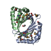
| ||||||||||||||||||||||||||||||
| 2 | 
| ||||||||||||||||||||||||||||||
| Unit cell |
| ||||||||||||||||||||||||||||||
| Noncrystallographic symmetry (NCS) | NCS domain:
NCS domain segments: Component-ID: 1 / Ens-ID: 1 / Beg auth comp-ID: GLU / Beg label comp-ID: GLU / End auth comp-ID: OHA / End label comp-ID: OHA / Refine code: 6 / Auth seq-ID: 3 - 300 / Label seq-ID: 4
| ||||||||||||||||||||||||||||||
| Details | SIZE EXCLUSION CHROMATOGRAPHY WITH STATIC LIGHT SCATTERING SUPPORTS THE ASSIGNMENT OF A DIMER AS A BIOLOGICALLY SIGNIFICANT OLIGOMERIZATION STATE. |
-Components
| #1: Protein | Mass: 12978.046 Da / Num. of mol.: 4 Source method: isolated from a genetically manipulated source Source: (gene. exp.) uncultured marine organism (environmental samples) Description: SYNTHETIC GENE: The gene product was based on JCVI_PEP_1096682647733 from the Sorcerer II Global Ocean Sampling experiment Production host:  #2: Chemical | ChemComp-OHA /   Mass: 270.408 Da / Num. of mol.: 4 / Source method: obtained synthetically / Formula: C16H30O3 Mass: 270.408 Da / Num. of mol.: 4 / Source method: obtained synthetically / Formula: C16H30O3#3: Chemical | ChemComp-EDO /   Mass: 62.068 Da / Num. of mol.: 11 / Source method: obtained synthetically / Formula: C2H6O2 Mass: 62.068 Da / Num. of mol.: 11 / Source method: obtained synthetically / Formula: C2H6O2#4: Water | ChemComp-HOH / |  Mass: 18.015 Da / Num. of mol.: 301 / Source method: isolated from a natural source / Formula: H2O Mass: 18.015 Da / Num. of mol.: 301 / Source method: isolated from a natural source / Formula: H2OHas protein modification | Y | |
|---|
-Experimental details
-Experiment
| Experiment | Method: X-RAY DIFFRACTION / Number of used crystals: 1 |
|---|
- Sample preparation
| Crystal | Density Matthews: 2.36 Å3/Da / Density % sol: 47.89 % |
|---|---|
| Crystal grow | Temperature: 277 K / Method: vapor diffusion, sitting drop, nanodrop / pH: 9 Details: 30.0% PEG-6000, 0.1M Bicine pH 9.0, VAPOR DIFFUSION, SITTING DROP, NANODROP, temperature 277K |
-Data collection
| Diffraction | Mean temperature: 100 K | ||||||||||||||||||||||||||||||||||||||||||||||||||||||||||||||||||||||||||||||||||||||||||||||||||||||||||||||||||||||||||||||||||||||||||||||||||||||||||||||||||||||||
|---|---|---|---|---|---|---|---|---|---|---|---|---|---|---|---|---|---|---|---|---|---|---|---|---|---|---|---|---|---|---|---|---|---|---|---|---|---|---|---|---|---|---|---|---|---|---|---|---|---|---|---|---|---|---|---|---|---|---|---|---|---|---|---|---|---|---|---|---|---|---|---|---|---|---|---|---|---|---|---|---|---|---|---|---|---|---|---|---|---|---|---|---|---|---|---|---|---|---|---|---|---|---|---|---|---|---|---|---|---|---|---|---|---|---|---|---|---|---|---|---|---|---|---|---|---|---|---|---|---|---|---|---|---|---|---|---|---|---|---|---|---|---|---|---|---|---|---|---|---|---|---|---|---|---|---|---|---|---|---|---|---|---|---|---|---|---|---|---|---|
| Diffraction source | Source: SYNCHROTRON / Site: SSRL  / Beamline: BL11-1 / Wavelength: 0.91837, 0.97944, 0.97972 / Beamline: BL11-1 / Wavelength: 0.91837, 0.97944, 0.97972 | ||||||||||||||||||||||||||||||||||||||||||||||||||||||||||||||||||||||||||||||||||||||||||||||||||||||||||||||||||||||||||||||||||||||||||||||||||||||||||||||||||||||||
| Detector | Type: MARMOSAIC 325 mm CCD / Detector: CCD / Date: Nov 19, 2006 / Details: Flat mirror (vertical focusing) | ||||||||||||||||||||||||||||||||||||||||||||||||||||||||||||||||||||||||||||||||||||||||||||||||||||||||||||||||||||||||||||||||||||||||||||||||||||||||||||||||||||||||
| Radiation | Monochromator: Single crystal Si(111) bent monochromator (horizontal focusing) Protocol: MAD / Monochromatic (M) / Laue (L): M / Scattering type: x-ray | ||||||||||||||||||||||||||||||||||||||||||||||||||||||||||||||||||||||||||||||||||||||||||||||||||||||||||||||||||||||||||||||||||||||||||||||||||||||||||||||||||||||||
| Radiation wavelength |
| ||||||||||||||||||||||||||||||||||||||||||||||||||||||||||||||||||||||||||||||||||||||||||||||||||||||||||||||||||||||||||||||||||||||||||||||||||||||||||||||||||||||||
| Reflection | Resolution: 1.8→29.086 Å / Num. obs: 42961 / % possible obs: 99.8 % / Redundancy: 3.6 % / Rmerge(I) obs: 0.072 / Rsym value: 0.072 / Net I/σ(I): 6.1 | ||||||||||||||||||||||||||||||||||||||||||||||||||||||||||||||||||||||||||||||||||||||||||||||||||||||||||||||||||||||||||||||||||||||||||||||||||||||||||||||||||||||||
| Reflection shell | Diffraction-ID: 1
|
-Phasing
| Phasing | Method: MAD |
|---|
- Processing
| Software |
| ||||||||||||||||||||||||||||||||||||||||||||||||||||||||||||||||||||||||||||||||||||||||||||||||||||||||||||||||||||||||||||||||||
|---|---|---|---|---|---|---|---|---|---|---|---|---|---|---|---|---|---|---|---|---|---|---|---|---|---|---|---|---|---|---|---|---|---|---|---|---|---|---|---|---|---|---|---|---|---|---|---|---|---|---|---|---|---|---|---|---|---|---|---|---|---|---|---|---|---|---|---|---|---|---|---|---|---|---|---|---|---|---|---|---|---|---|---|---|---|---|---|---|---|---|---|---|---|---|---|---|---|---|---|---|---|---|---|---|---|---|---|---|---|---|---|---|---|---|---|---|---|---|---|---|---|---|---|---|---|---|---|---|---|---|---|
| Refinement | Method to determine structure: MAD / Resolution: 1.85→29.086 Å / Cor.coef. Fo:Fc: 0.963 / Cor.coef. Fo:Fc free: 0.952 / SU B: 9.082 / SU ML: 0.132 / TLS residual ADP flag: LIKELY RESIDUAL / Cross valid method: THROUGHOUT / σ(F): 0 / ESU R: 0.151 / ESU R Free: 0.137 Stereochemistry target values: MAXIMUM LIKELIHOOD WITH PHASES Details: (1). HYDROGENS HAVE BEEN ADDED IN RIDING POSITIONS. (2). ATOM RECORD CONTAINS RESIDUAL B FACTORS ONLY. (3). A MET-INHIBITION PROTOCOL WAS USED FOR SELENOMETHIONINE INCORPORATION DURING ...Details: (1). HYDROGENS HAVE BEEN ADDED IN RIDING POSITIONS. (2). ATOM RECORD CONTAINS RESIDUAL B FACTORS ONLY. (3). A MET-INHIBITION PROTOCOL WAS USED FOR SELENOMETHIONINE INCORPORATION DURING PROTEIN EXPRESSION. THE OCCUPANCY OF THE SE ATOMS IN THE MSE RESIDUES WAS REDUCED TO 0.75 TO ACCOUNT FOR THE REDUCED SCATTERING POWER DUE TO PARTIAL S-MET INCORPORATION. (4). RESIDUES 85-88 IN A SUBUNIT ARE DISORDERED. THE GEOMETRY IS POOR IN THIS REGION. (5). 10-KETO PALMITIC ACIDS (10-OXO-HEXADECANOIC ACID; OHA) WERE MODELLED IN THE ACTIVE SITE POCKETS. THE CARBOXYL GROUPS OF THE HEAD GROUPS OF THE FATTY ACIDS INTERACT WITH THE SIDE CHAINS OF SER 8, TYR 66, ARG 102 AND TYR 64. THE OHA ASSOCIATED WITH CHAIN C HAS THE BEST DENSITY. THE ASSIGNMENT OF THE FATTY ACIDS WAS BASED ON ELECTRON DENSITY AND THE STRUCTURAL SIMILARITY WITH OTHER MONOOXYGENASES. THEY COULD BE OTHER SIMILAR LIPIDS.
| ||||||||||||||||||||||||||||||||||||||||||||||||||||||||||||||||||||||||||||||||||||||||||||||||||||||||||||||||||||||||||||||||||
| Solvent computation | Ion probe radii: 0.8 Å / Shrinkage radii: 0.8 Å / VDW probe radii: 1.2 Å / Solvent model: MASK | ||||||||||||||||||||||||||||||||||||||||||||||||||||||||||||||||||||||||||||||||||||||||||||||||||||||||||||||||||||||||||||||||||
| Displacement parameters | Biso mean: 11.009 Å2
| ||||||||||||||||||||||||||||||||||||||||||||||||||||||||||||||||||||||||||||||||||||||||||||||||||||||||||||||||||||||||||||||||||
| Refinement step | Cycle: LAST / Resolution: 1.85→29.086 Å
| ||||||||||||||||||||||||||||||||||||||||||||||||||||||||||||||||||||||||||||||||||||||||||||||||||||||||||||||||||||||||||||||||||
| Refine LS restraints |
| ||||||||||||||||||||||||||||||||||||||||||||||||||||||||||||||||||||||||||||||||||||||||||||||||||||||||||||||||||||||||||||||||||
| Refine LS restraints NCS | Ens-ID: 1 / Number: 1265 / Refine-ID: X-RAY DIFFRACTION
| ||||||||||||||||||||||||||||||||||||||||||||||||||||||||||||||||||||||||||||||||||||||||||||||||||||||||||||||||||||||||||||||||||
| LS refinement shell | Resolution: 1.85→1.898 Å / Total num. of bins used: 20
| ||||||||||||||||||||||||||||||||||||||||||||||||||||||||||||||||||||||||||||||||||||||||||||||||||||||||||||||||||||||||||||||||||
| Refinement TLS params. | Method: refined / Refine-ID: X-RAY DIFFRACTION
| ||||||||||||||||||||||||||||||||||||||||||||||||||||||||||||||||||||||||||||||||||||||||||||||||||||||||||||||||||||||||||||||||||
| Refinement TLS group |
|
