 Movie
Movie Controller

[English] 日本語
 Yorodumi
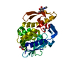
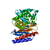
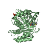
- PDB-3bfd: Crystal Structure of the Class A beta-lactamase SED-G238C mutant ... -
+ Open data

- Basic information
| Entry | Database: PDB / ID: 3bfd | ||||||
|---|---|---|---|---|---|---|---|
| Title | Crystal Structure of the Class A beta-lactamase SED-G238C mutant from Citrobacter sedlakii | ||||||
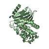
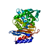
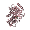
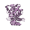
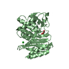
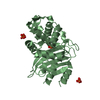
 Components Components | Class A beta-lactamase Sed1 | ||||||
 Keywords Keywords | HYDROLASE / BETA-LACTAMASE / CLASS A / SED-G238C MUTANT | ||||||
| Function / homology |  Function and homology information Function and homology informationbeta-lactam antibiotic catabolic process / beta-lactamase activity / beta-lactamase / response to antibiotic Similarity search - Function | ||||||
| Biological species |  Citrobacter sedlakii (bacteria) Citrobacter sedlakii (bacteria) | ||||||
| Method |  X-RAY DIFFRACTION / SYNCHROTRON / MOLECULAR REPLACEMENT / Resolution: 2 Å X-RAY DIFFRACTION / SYNCHROTRON / MOLECULAR REPLACEMENT / Resolution: 2 Å | ||||||
 Authors Authors | Pernot, L. / Petrella, S. / Sougakoff, W. | ||||||
 Citation Citation | Journal: To be Published Title: acyl-intermediate structures of the class A beta-lactamase SED-G238C Authors: Pernot, L. / Petrella, S. / Sougakoff, W. #1: Journal: Acta Crystallogr.,Sect.D / Year: 2004 Title: Crystallization and preliminary X-ray diffraction study of the class A beta-lactamase SED-1 and its mutant SED-G238C from Citrobacter sedlakii Authors: Petrella, S. / Pernot, L. / Sougakoff, W. | ||||||
| History |
|
- Structure visualization
| Structure viewer | Molecule: MolmilJmol/JSmol |
|---|
- Downloads & links
-Download
| PDBx/mmCIF format | 3bfd.cif.gz | 228 KB | Display | PDBx/mmCIF format |
|---|---|---|---|---|
| PDB format | pdb3bfd.ent.gz | 181.2 KB | Display | PDB format |
| PDBx/mmJSON format | 3bfd.json.gz | Tree view | PDBx/mmJSON format | |
| Others |  Other downloads Other downloads |
-Validation report
| Arichive directory | https://data.pdbj.org/pub/pdb/validation_reports/bf/3bfdftp://data.pdbj.org/pub/pdb/validation_reports/bf/3bfd | HTTPS FTP |
|---|
-Related structure data
| Related structure data |  3bfeC 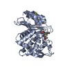 3bffC  3bfgC  1bzaS C: citing same article ( S: Starting model for refinement |
|---|---|
| Similar structure data |


-Links
 PDBj
PDBj


- Assembly
| Deposited unit | 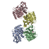
| ||||||||
|---|---|---|---|---|---|---|---|---|---|
| 1 | 
| ||||||||
| 2 | 
| ||||||||
| 3 | 
| ||||||||
| 4 | 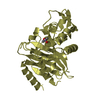
| ||||||||
| Unit cell |
|
-Components
| #1: Protein | Mass: 28445.307 Da / Num. of mol.: 4 / Fragment: SED-G238C, UNP residues 33-295 / Mutation: G238C Source method: isolated from a genetically manipulated source Source: (gene. exp.) Citrobacter sedlakii (bacteria) / Strain: 2596 / Gene: bla-SED-1 / Plasmid: pET29a / Species (production host): Escherichia coli / Production host: #2: Chemical | ChemComp-CAC /   Mass: 136.989 Da / Num. of mol.: 4 / Source method: obtained synthetically / Formula: C2H6AsO2 Mass: 136.989 Da / Num. of mol.: 4 / Source method: obtained synthetically / Formula: C2H6AsO2#3: Water | ChemComp-HOH / |  Mass: 18.015 Da / Num. of mol.: 1019 / Source method: isolated from a natural source / Formula: H2O Mass: 18.015 Da / Num. of mol.: 1019 / Source method: isolated from a natural source / Formula: H2OHas protein modification | Y | Sequence details | THIS COORDINATES IS USED NON-SEQUENTIAL RESIDUE NUMBERING. THREE NUMBERS, 58, 239, AND 253 WERE ...THIS COORDINATE | |
|---|
-Experimental details
-Experiment
| Experiment | Method: X-RAY DIFFRACTION / Number of used crystals: 1 |
|---|
- Sample preparation
| Crystal | Density Matthews: 2.66 Å3/Da / Density % sol: 53.8 % |
|---|---|
| Crystal grow | Temperature: 291 K / Method: vapor diffusion, hanging drop / pH: 6.5 Details: 35% PEG MME 2000, 200mM KSCN, 100mM sodium cacodylate pH 6.5, VAPOR DIFFUSION, HANGING DROP, temperature 291K |
-Data collection
| Diffraction | Mean temperature: 100 K |
|---|---|
| Diffraction source | Source: SYNCHROTRON / Site: ESRF  / Beamline: BM14 / Wavelength: 0.97887 Å / Beamline: BM14 / Wavelength: 0.97887 Å |
| Detector | Type: MARRESEARCH / Detector: CCD / Date: Jul 18, 2002 |
| Radiation | Monochromator: Si (1 1 1) / Protocol: SINGLE WAVELENGTH / Monochromatic (M) / Laue (L): M / Scattering type: x-ray |
| Radiation wavelength | Wavelength: 0.97887 Å / Relative weight: 1 |
| Reflection | Resolution: 2→16.7 Å / Num. all: 80352 / Num. obs: 80352 / % possible obs: 99.3 % / Redundancy: 3.5 % / Rsym value: 0.102 / Net I/σ(I): 8.7 |
| Reflection shell | Resolution: 2→2.11 Å / Redundancy: 2.3 % / Mean I/σ(I) obs: 2 / Num. unique all: 11334 / Rsym value: 0.33 / % possible all: 96.4 |
- Processing
| Software |
| |||||||||||||||||||||||||
|---|---|---|---|---|---|---|---|---|---|---|---|---|---|---|---|---|---|---|---|---|---|---|---|---|---|---|
| Refinement | Method to determine structure: MOLECULAR REPLACEMENT Starting model: PDB ENTRY 1BZA Resolution: 2→16.7 Å / Isotropic thermal model: restrained / Cross valid method: THROUGHOUT / Stereochemistry target values: Engh & Huber
| |||||||||||||||||||||||||
| Displacement parameters | Biso mean: 16.92 Å2
| |||||||||||||||||||||||||
| Refine analyze |
| |||||||||||||||||||||||||
| Refinement step | Cycle: LAST / Resolution: 2→16.7 Å
| |||||||||||||||||||||||||
| Refine LS restraints |
| |||||||||||||||||||||||||
| LS refinement shell | Resolution: 2→2.13 Å / Rfactor Rfree error: 0.008
|
