 Movie
Movie Controller

[English] 日本語
 Yorodumi
- PDB-1o03: Structure of Pentavalent Phosphorous Intermediate of an Enzyme Ca... -
+ Open data

- Basic information
| Entry | Database: PDB / ID: 1o03 | ||||||
|---|---|---|---|---|---|---|---|
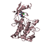
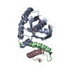
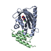
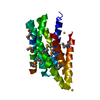
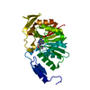
| Title | Structure of Pentavalent Phosphorous Intermediate of an Enzyme Catalyzed Phosphoryl transfer Reaction observed on cocrystallization with Glucose 6-phosphate | ||||||
 Components Components | beta-phosphoglucomutase | ||||||
 Keywords Keywords | ISOMERASE / Haloacid dehalogenase superfamily / phosphotransferase / pentavalent phosphate intermediate | ||||||
| Function / homology |  Function and homology information Function and homology informationbeta-phosphoglucomutase / beta-phosphoglucomutase activity / carbohydrate metabolic process / magnesium ion binding / cytoplasm Similarity search - Function | ||||||
| Biological species |  Lactococcus lactis (lactic acid bacteria) Lactococcus lactis (lactic acid bacteria) | ||||||
| Method |  X-RAY DIFFRACTION / SYNCHROTRON / MOLECULAR REPLACEMENT / Resolution: 1.4 Å X-RAY DIFFRACTION / SYNCHROTRON / MOLECULAR REPLACEMENT / Resolution: 1.4 Å | ||||||
 Authors Authors | Lahiri, S.D. / Zhang, G. / Dunaway-Mariano, D. / Allen, K.N. | ||||||
 Citation Citation | Journal: Science / Year: 2003 Title: The pentacovalent phosphorus intermediate of a phosphoryl transfer reaction. Authors: Lahiri, S.D. / Zhang, G. / Dunaway-Mariano, D. / Allen, K.N. | ||||||
| History |
|
- Structure visualization
| Structure viewer | Molecule: MolmilJmol/JSmol |
|---|
- Downloads & links
-Download
| PDBx/mmCIF format | 1o03.cif.gz | 67.9 KB | Display | PDBx/mmCIF format |
|---|---|---|---|---|
| PDB format | pdb1o03.ent.gz | 48.4 KB | Display | PDB format |
| PDBx/mmJSON format | 1o03.json.gz | Tree view | PDBx/mmJSON format | |
| Others |  Other downloads Other downloads |
-Validation report
| Arichive directory | https://data.pdbj.org/pub/pdb/validation_reports/o0/1o03ftp://data.pdbj.org/pub/pdb/validation_reports/o0/1o03 | HTTPS FTP |
|---|
-Related structure data


-Links
 PDBj
PDBj

- Assembly
| Deposited unit | 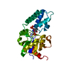
| ||||||||
|---|---|---|---|---|---|---|---|---|---|
| 1 |
| ||||||||
| Unit cell |
|
-Components
| #1: Protein | Mass: 24239.594 Da / Num. of mol.: 1 Source method: isolated from a genetically manipulated source Source: (gene. exp.) Lactococcus lactis (lactic acid bacteria)Species (production host): Escherichia coli / Production host: |
|---|---|
| #2: Sugar | ChemComp-G16 /   Type: D-saccharide / Mass: 339.108 Da / Num. of mol.: 1 Type: D-saccharide / Mass: 339.108 Da / Num. of mol.: 1Source method: isolated from a genetically manipulated source Formula: C6H13O12P2 |
| #3: Chemical | ChemComp-MG /   Mass: 24.305 Da / Num. of mol.: 1 / Source method: obtained synthetically / Formula: Mg Mass: 24.305 Da / Num. of mol.: 1 / Source method: obtained synthetically / Formula: Mg |
| #4: Water | ChemComp-HOH /  Mass: 18.015 Da / Num. of mol.: 402 / Source method: isolated from a natural source / Formula: H2O Mass: 18.015 Da / Num. of mol.: 402 / Source method: isolated from a natural source / Formula: H2O |
-Experimental details
-Experiment
| Experiment | Method: X-RAY DIFFRACTION / Number of used crystals: 1 |
|---|
- Sample preparation
| Crystal | Density Matthews: 1.72 Å3/Da / Density % sol: 27.99 % | ||||||||||||||||||||||||||||||||||||||||||
|---|---|---|---|---|---|---|---|---|---|---|---|---|---|---|---|---|---|---|---|---|---|---|---|---|---|---|---|---|---|---|---|---|---|---|---|---|---|---|---|---|---|---|---|
| Crystal grow | Temperature: 291 K / Method: vapor diffusion, hanging drop / pH: 6.9 Details: 16% PEG 3350, 0.1M Ammonium Fluoride, 4mM b-D-Glucose6phosphate, pH 6.9, VAPOR DIFFUSION, HANGING DROP, temperature 291K | ||||||||||||||||||||||||||||||||||||||||||
| Crystal grow | *PLUS Temperature: 18 ℃ | ||||||||||||||||||||||||||||||||||||||||||
| Components of the solutions | *PLUS
|
-Data collection
| Diffraction | Mean temperature: 93 K |
|---|---|
| Diffraction source | Source: SYNCHROTRON / Site: APS  / Beamline: 14-BM-C / Wavelength: 0.9 Å / Beamline: 14-BM-C / Wavelength: 0.9 Å |
| Detector | Type: ADSC QUANTUM 4 / Detector: CCD / Date: Feb 22, 2002 / Details: mirrors |
| Radiation | Monochromator: Bent Ge(111) / Protocol: SINGLE WAVELENGTH / Monochromatic (M) / Laue (L): M / Scattering type: x-ray |
| Radiation wavelength | Wavelength: 0.9 Å / Relative weight: 1 |
| Reflection | Resolution: 1.4→35.14 Å / Num. obs: 41217 / % possible obs: 96.7 % / Observed criterion σ(F): 0 / Observed criterion σ(I): 0 / Redundancy: 14.8 % / Biso Wilson estimate: 14.3 Å2 / Rmerge(I) obs: 0.1 / Rsym value: 0.09 / Net I/σ(I): 34.9 |
| Reflection shell | Resolution: 1.4→1.45 Å / Redundancy: 5 % / Rmerge(I) obs: 0.17 / Mean I/σ(I) obs: 15.7 / Rsym value: 0.19 / % possible all: 88 |
| Reflection | *PLUS Lowest resolution: 9999 Å / Num. obs: 66302 / % possible obs: 98 % / Num. measured all: 983130 / Rmerge(I) obs: 0.073 |
| Reflection shell | *PLUS % possible obs: 88.1 % / Rmerge(I) obs: 0.13 |
- Processing
| Software |
| ||||||||||||||||||||||||||||||||||||
|---|---|---|---|---|---|---|---|---|---|---|---|---|---|---|---|---|---|---|---|---|---|---|---|---|---|---|---|---|---|---|---|---|---|---|---|---|---|
| Refinement | Method to determine structure: MOLECULAR REPLACEMENT Starting model: SeMeth MAD model of the same protein to 1.7 angstrom Resolution: 1.4→35.14 Å / Isotropic thermal model: Isotropic / Cross valid method: THROUGHOUT / σ(F): 0 / σ(I): 0 / Stereochemistry target values: Engh & Huber / Details: used mlf refinement
| ||||||||||||||||||||||||||||||||||||
| Displacement parameters | Biso mean: 13.7 Å2
| ||||||||||||||||||||||||||||||||||||
| Refine analyze |
| ||||||||||||||||||||||||||||||||||||
| Refinement step | Cycle: LAST / Resolution: 1.4→35.14 Å
| ||||||||||||||||||||||||||||||||||||
| Refine LS restraints |
| ||||||||||||||||||||||||||||||||||||
| LS refinement shell | Resolution: 1.4→1.49 Å / Rfactor Rfree error: 0.012
| ||||||||||||||||||||||||||||||||||||
| Xplor file |
| ||||||||||||||||||||||||||||||||||||
| Refinement | *PLUS Lowest resolution: 100 Å / % reflection Rfree: 10 % / Rfactor Rfree: 0.1944 / Rfactor Rwork: 0.1702 | ||||||||||||||||||||||||||||||||||||
| Solvent computation | *PLUS | ||||||||||||||||||||||||||||||||||||
| Displacement parameters | *PLUS | ||||||||||||||||||||||||||||||||||||
| Refine LS restraints | *PLUS
| ||||||||||||||||||||||||||||||||||||
| LS refinement shell | *PLUS Lowest resolution: 1.45 Å |
