 Movie
Movie Controller

[English] 日本語
 Yorodumi
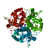
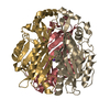
- PDB-7jmv: Crystal structure of the pea pathogenicity protein 2 from Madurel... -
+ Open data

- Basic information
| Entry | Database: PDB / ID: 7jmv | ||||||
|---|---|---|---|---|---|---|---|
| Title | Crystal structure of the pea pathogenicity protein 2 from Madurella mycetomatis complexed with 4-nitrocatechol | ||||||
 Components Components | Pea pathogenicity protein 2 | ||||||
 Keywords Keywords | LYASE / Gallic acid decarboxylase | ||||||
| Function / homology | SnoaL-like domain / SnoaL-like domain / WW/rsp5/WWP domain profile. / WW domain / NTF2-like domain superfamily / metal ion binding / 4-NITROCATECHOL / : / Pea pathogenicity protein 2 Function and homology information Function and homology information | ||||||
| Biological species |  Madurella mycetomatis (fungus) Madurella mycetomatis (fungus) | ||||||
| Method |  X-RAY DIFFRACTION / SYNCHROTRON / MOLECULAR REPLACEMENT / Resolution: 1.57 Å X-RAY DIFFRACTION / SYNCHROTRON / MOLECULAR REPLACEMENT / Resolution: 1.57 Å | ||||||
 Authors Authors | Zeug, M. / Markovic, N. / Iancu, C.V. / Tripp, J. / Oreb, M. / Choe, J. | ||||||
 Citation Citation | Journal: Sci Rep / Year: 2021 Title: Crystal structures of non-oxidative decarboxylases reveal a new mechanism of action with a catalytic dyad and structural twists. Authors: Zeug, M. / Markovic, N. / Iancu, C.V. / Tripp, J. / Oreb, M. / Choe, J.Y. | ||||||
| History |
|
- Structure visualization
| Structure viewer | Molecule: MolmilJmol/JSmol |
|---|
- Downloads & links
-Download
| PDBx/mmCIF format | 7jmv.cif.gz | 101 KB | Display | PDBx/mmCIF format |
|---|---|---|---|---|
| PDB format | pdb7jmv.ent.gz | 76.5 KB | Display | PDB format |
| PDBx/mmJSON format | 7jmv.json.gz | Tree view | PDBx/mmJSON format | |
| Others |  Other downloads Other downloads |
-Validation report
| Arichive directory | https://data.pdbj.org/pub/pdb/validation_reports/jm/7jmvftp://data.pdbj.org/pub/pdb/validation_reports/jm/7jmv | HTTPS FTP |
|---|
-Related structure data
| Related structure data | 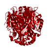 6w54C  7jmrSC 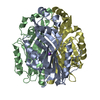 7kd9C S: Starting model for refinement C: citing same article ( |
|---|---|
| Similar structure data |


-Links
 PDBj
PDBj

- Assembly
| Deposited unit | 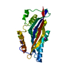
| |||||||||||||||||||||
|---|---|---|---|---|---|---|---|---|---|---|---|---|---|---|---|---|---|---|---|---|---|---|
| 1 | 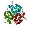
| |||||||||||||||||||||
| Unit cell |
| |||||||||||||||||||||
| Components on special symmetry positions |
|
-Components
| #1: Protein | Mass: 27406.799 Da / Num. of mol.: 1 Source method: isolated from a genetically manipulated source Source: (gene. exp.) Madurella mycetomatis (fungus) / Gene: MMYC01_201259, MMYC01_203104 / Production host:  | ||||
|---|---|---|---|---|---|
| #2: Chemical | ChemComp-4NC / 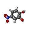  Mass: 155.108 Da / Num. of mol.: 1 / Source method: obtained synthetically / Formula: C6H5NO4 / Feature type: SUBJECT OF INVESTIGATION Mass: 155.108 Da / Num. of mol.: 1 / Source method: obtained synthetically / Formula: C6H5NO4 / Feature type: SUBJECT OF INVESTIGATION | ||||
| #3: Chemical | ChemComp-K /   Mass: 39.098 Da / Num. of mol.: 1 / Source method: obtained synthetically / Formula: K / Feature type: SUBJECT OF INVESTIGATION Mass: 39.098 Da / Num. of mol.: 1 / Source method: obtained synthetically / Formula: K / Feature type: SUBJECT OF INVESTIGATION | ||||
| #4: Chemical |   Mass: 40.078 Da / Num. of mol.: 2 / Source method: obtained synthetically / Formula: Ca Mass: 40.078 Da / Num. of mol.: 2 / Source method: obtained synthetically / Formula: Ca#5: Water | ChemComp-HOH / |  Mass: 18.015 Da / Num. of mol.: 163 / Source method: isolated from a natural source / Formula: H2O Mass: 18.015 Da / Num. of mol.: 163 / Source method: isolated from a natural source / Formula: H2OHas ligand of interest | Y | |
-Experimental details
-Experiment
| Experiment | Method: X-RAY DIFFRACTION / Number of used crystals: 1 |
|---|
- Sample preparation
| Crystal | Density Matthews: 3.2 Å3/Da / Density % sol: 61.54 % |
|---|---|
| Crystal grow | Temperature: 295 K / Method: vapor diffusion, hanging drop / Details: 15 % PEG 3350, 0.2 M Calcium Chloride, 0.1 M MES / PH range: 5.5 |
-Data collection
| Diffraction | Mean temperature: 100 K / Serial crystal experiment: N |
|---|---|
| Diffraction source | Source: SYNCHROTRON / Site: APS  / Beamline: 23-ID-D / Wavelength: 0.97934 Å / Beamline: 23-ID-D / Wavelength: 0.97934 Å |
| Detector | Type: DECTRIS PILATUS3 6M / Detector: PIXEL / Date: Feb 4, 2017 |
| Radiation | Protocol: SINGLE WAVELENGTH / Monochromatic (M) / Laue (L): M / Scattering type: x-ray |
| Radiation wavelength | Wavelength: 0.97934 Å / Relative weight: 1 |
| Reflection | Resolution: 1.57→42.76 Å / Num. obs: 49087 / % possible obs: 99.9 % / Redundancy: 9.8 % / Rsym value: 0.08 / Net I/σ(I): 41.4 |
| Reflection shell | Resolution: 1.57→1.6 Å / Num. unique obs: 3019 / Rsym value: 0.894 |
- Processing
| Software |
| |||||||||||||||||||||||||||||||||||||||||||||||||||||||||||||||||||||||||||||||||||||||||||||||||||||||||||||||||||||||
|---|---|---|---|---|---|---|---|---|---|---|---|---|---|---|---|---|---|---|---|---|---|---|---|---|---|---|---|---|---|---|---|---|---|---|---|---|---|---|---|---|---|---|---|---|---|---|---|---|---|---|---|---|---|---|---|---|---|---|---|---|---|---|---|---|---|---|---|---|---|---|---|---|---|---|---|---|---|---|---|---|---|---|---|---|---|---|---|---|---|---|---|---|---|---|---|---|---|---|---|---|---|---|---|---|---|---|---|---|---|---|---|---|---|---|---|---|---|---|---|---|
| Refinement | Method to determine structure: MOLECULAR REPLACEMENT Starting model: 7JMR Resolution: 1.57→42.76 Å / SU ML: 0.17 / Cross valid method: THROUGHOUT / σ(F): 1.38 / Phase error: 18.01 / Stereochemistry target values: ML
| |||||||||||||||||||||||||||||||||||||||||||||||||||||||||||||||||||||||||||||||||||||||||||||||||||||||||||||||||||||||
| Solvent computation | Shrinkage radii: 0.9 Å / VDW probe radii: 1.11 Å / Solvent model: FLAT BULK SOLVENT MODEL | |||||||||||||||||||||||||||||||||||||||||||||||||||||||||||||||||||||||||||||||||||||||||||||||||||||||||||||||||||||||
| Displacement parameters | Biso max: 94.14 Å2 / Biso mean: 26.764 Å2 / Biso min: 8.58 Å2 | |||||||||||||||||||||||||||||||||||||||||||||||||||||||||||||||||||||||||||||||||||||||||||||||||||||||||||||||||||||||
| Refinement step | Cycle: final / Resolution: 1.57→42.76 Å
| |||||||||||||||||||||||||||||||||||||||||||||||||||||||||||||||||||||||||||||||||||||||||||||||||||||||||||||||||||||||
| Refine LS restraints |
| |||||||||||||||||||||||||||||||||||||||||||||||||||||||||||||||||||||||||||||||||||||||||||||||||||||||||||||||||||||||
| LS refinement shell | Refine-ID: X-RAY DIFFRACTION / Rfactor Rfree error: 0 / Total num. of bins used: 16
|
