 Movie
Movie Controller


+ Open data

- Basic information
| Entry | Database: PDB / ID: 1nwp | ||||||
|---|---|---|---|---|---|---|---|
| Title | CRYSTALLOGRAPHIC STUDY OF AZURIN FROM PSEUDOMONAS PUTIDA | ||||||
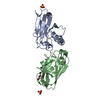
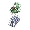
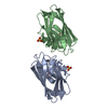
 Components Components | AZURIN | ||||||
 Keywords Keywords | ELECTRON TRANSPORT / CUPREDOXIN / ELECTRON TRANSFER | ||||||
| Function / homology |  Function and homology information Function and homology information | ||||||
| Biological species |  Pseudomonas putida (bacteria) Pseudomonas putida (bacteria) | ||||||
| Method |  X-RAY DIFFRACTION / MOLECULAR REPLACEMENT / Resolution: 1.6 Å X-RAY DIFFRACTION / MOLECULAR REPLACEMENT / Resolution: 1.6 Å | ||||||
 Authors Authors | Mathews, F.S. / Chen, Z.-W. | ||||||
 Citation Citation | Journal: Acta Crystallogr.,Sect.D / Year: 1998 Title: Crystallographic study of azurin from Pseudomonas putida. Authors: Chen, Z.W. / Barber, M.J. / McIntire, W.S. / Mathews, F.S. #1: Journal: Arch.Biochem.Biophys. / Year: 1993Title: The Amino Acid Sequence of Pseudomonas Putida Azurin Authors: Barber, M.J. / Trimboli, A.J. / Mcintire, W.S. | ||||||
| History |
|
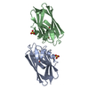
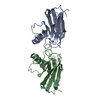
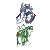
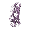
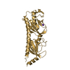
- Structure visualization
| Structure viewer | Molecule: MolmilJmol/JSmol |
|---|
- Downloads & links
-Download
| PDBx/mmCIF format | 1nwp.cif.gz | 65.5 KB | Display | PDBx/mmCIF format |
|---|---|---|---|---|
| PDB format | pdb1nwp.ent.gz | 47.7 KB | Display | PDB format |
| PDBx/mmJSON format | 1nwp.json.gz | Tree view | PDBx/mmJSON format | |
| Others |  Other downloads Other downloads |
-Validation report
| Arichive directory | https://data.pdbj.org/pub/pdb/validation_reports/nw/1nwpftp://data.pdbj.org/pub/pdb/validation_reports/nw/1nwp | HTTPS FTP |
|---|
-Related structure data
| Related structure data |  1nwoC 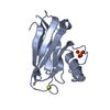 2azaS S: Starting model for refinement C: citing same article ( |
|---|---|
| Similar structure data |


-Links
 PDBj
PDBj- Assembly
| Deposited unit | 
| ||||||||
|---|---|---|---|---|---|---|---|---|---|
| 1 |
| ||||||||
| 2 | 
| ||||||||
| 3 | 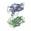
| ||||||||
| Unit cell |
|
-Components
| #1: Protein | Mass: 13737.709 Da / Num. of mol.: 2 / Source method: isolated from a natural source / Source: (natural) Pseudomonas putida (bacteria) / Strain: NCIB 9869 / References: UniProt: P34097#2: Chemical |   Mass: 63.546 Da / Num. of mol.: 2 / Source method: obtained synthetically / Formula: Cu Mass: 63.546 Da / Num. of mol.: 2 / Source method: obtained synthetically / Formula: Cu#3: Chemical | ChemComp-ZN /   Mass: 65.409 Da / Num. of mol.: 4 / Source method: obtained synthetically / Formula: Zn Mass: 65.409 Da / Num. of mol.: 4 / Source method: obtained synthetically / Formula: Zn#4: Water | ChemComp-HOH / |  Mass: 18.015 Da / Num. of mol.: 222 / Source method: isolated from a natural source / Formula: H2O Mass: 18.015 Da / Num. of mol.: 222 / Source method: isolated from a natural source / Formula: H2OHas protein modification | Y | |
|---|
-Experimental details
-Experiment
| Experiment | Method: X-RAY DIFFRACTION / Number of used crystals: 1 |
|---|
- Sample preparation
| Crystal | Density Matthews: 2 Å3/Da / Density % sol: 38.5 % | ||||||||||||||||||||||||||||||||||||||||||||||||||||||
|---|---|---|---|---|---|---|---|---|---|---|---|---|---|---|---|---|---|---|---|---|---|---|---|---|---|---|---|---|---|---|---|---|---|---|---|---|---|---|---|---|---|---|---|---|---|---|---|---|---|---|---|---|---|---|---|
| Crystal grow | Temperature: 277 K / Method: vapor diffusion, hanging drop / pH: 7 Details: HANGING DROP METHOD AT 4 C BY MIXING 5 MICROLITER PROTEIN AT 10- 15MG PER ML WITH PEG8000 SOLUTION CONTAINING 5MM TRIS-HCL BUFFER, PH 5 MICROLITER 30-36% 6.5-7.5 ,100MM NACL AND 180MM ZINC ...Details: HANGING DROP METHOD AT 4 C BY MIXING 5 MICROLITER PROTEIN AT 10- 15MG PER ML WITH PEG8000 SOLUTION CONTAINING 5MM TRIS-HCL BUFFER, PH 5 MICROLITER 30-36% 6.5-7.5 ,100MM NACL AND 180MM ZINC ACETATE., pH 7.0, vapor diffusion - hanging drop, temperature 277K PH range: 6.5-7.5 | ||||||||||||||||||||||||||||||||||||||||||||||||||||||
| Crystal grow | *PLUS Temperature: 277 K / Method: vapor diffusion, hanging drop / PH range low: 7.5 / PH range high: 6.5 | ||||||||||||||||||||||||||||||||||||||||||||||||||||||
| Components of the solutions | *PLUS
|
-Data collection
| Diffraction | Mean temperature: 298 K |
|---|---|
| Diffraction source | Source: ROTATING ANODE / Type: RIGAKU RUH2R / Wavelength: 1.5418 |
| Detector | Type: XUONG-HAMLIN MULTIWIRE / Detector: AREA DETECTOR / Date: Apr 1, 1992 / Details: CU KA RADIATION |
| Radiation | Monochromator: GRAPHITE(002) / Monochromatic (M) / Laue (L): M / Scattering type: x-ray |
| Radiation wavelength | Wavelength: 1.5418 Å / Relative weight: 1 |
| Reflection | Resolution: 1.6→20 Å / Num. obs: 27846 / % possible obs: 95.4 % / Observed criterion σ(I): 0 / Redundancy: 8.7 % / Biso Wilson estimate: 14 Å2 / Rmerge(I) obs: 0.055 / Net I/σ(I): 10.7 |
| Reflection shell | Resolution: 1.6→1.72 Å / Redundancy: 3.2 % / Rmerge(I) obs: 0.218 / Mean I/σ(I) obs: 1.9 / % possible all: 78 |
| Reflection | *PLUS Num. measured all: 241088 |
- Processing
| Software |
| ||||||||||||||||||||||||||||||||||||||||||||||||||||||||||||
|---|---|---|---|---|---|---|---|---|---|---|---|---|---|---|---|---|---|---|---|---|---|---|---|---|---|---|---|---|---|---|---|---|---|---|---|---|---|---|---|---|---|---|---|---|---|---|---|---|---|---|---|---|---|---|---|---|---|---|---|---|---|
| Refinement | Method to determine structure: MOLECULAR REPLACEMENT Starting model: AZURIN FROM ALCALIGENES DENITRIFICANS (ENTRY NO. 2AZA) Resolution: 1.6→10 Å / σ(F): 1.5
| ||||||||||||||||||||||||||||||||||||||||||||||||||||||||||||
| Displacement parameters | Biso mean: 18 Å2 | ||||||||||||||||||||||||||||||||||||||||||||||||||||||||||||
| Refinement step | Cycle: LAST / Resolution: 1.6→10 Å
| ||||||||||||||||||||||||||||||||||||||||||||||||||||||||||||
| Refine LS restraints |
| ||||||||||||||||||||||||||||||||||||||||||||||||||||||||||||
| LS refinement shell | Resolution: 1.6→1.67 Å / Total num. of bins used: 8
| ||||||||||||||||||||||||||||||||||||||||||||||||||||||||||||
| Software | *PLUS Name: X-PLOR / Version: 3.1 / Classification: refinement | ||||||||||||||||||||||||||||||||||||||||||||||||||||||||||||
| Refine LS restraints | *PLUS
|
