 Movie
Movie Controller

[English] 日本語
 Yorodumi
- PDB-1nqk: Structural Genomics, Crystal structure of Alkanesulfonate monooxy... -
+ Open data

- Basic information
| Entry | Database: PDB / ID: 1nqk | ||||||
|---|---|---|---|---|---|---|---|
| Title | Structural Genomics, Crystal structure of Alkanesulfonate monooxygenase | ||||||
 Components Components | Alkanesulfonate monooxygenase | ||||||
 Keywords Keywords | OXIDOREDUCTASE / STRUCTURAL GENOMICS / BETA BARREL / PSI / Protein Structure Initiative / Midwest Center for Structural Genomics / MCSG | ||||||
| Function / homology |  Function and homology information Function and homology informationalkanesulfonate monooxygenase complex / alkanesulfonate monooxygenase / cellular response to sulfur starvation / alkanesulfonate monooxygenase activity / alkanesulfonate catabolic process / response to heat / protein homotetramerization / identical protein binding Similarity search - Function | ||||||
| Biological species |  | ||||||
| Method |  X-RAY DIFFRACTION / SYNCHROTRON / MAD / Resolution: 2.2 Å X-RAY DIFFRACTION / SYNCHROTRON / MAD / Resolution: 2.2 Å | ||||||
 Authors Authors | Zhang, R. / Skarina, T. / Savchenko, A. / Edwards, A. / Joachimiak, A. / Midwest Center for Structural Genomics (MCSG) | ||||||
 Citation Citation | Journal: To be Published Title: The crystal structure of the protein Alkanesulfonate monooxygenase from E. Coli Authors: Zhang, R. / Skarina, T. / Savchenko, A. / Edwards, A. / Joachimiak, A. | ||||||
| History |
|
- Structure visualization
| Structure viewer | Molecule: MolmilJmol/JSmol |
|---|
- Downloads & links
-Download
| PDBx/mmCIF format | 1nqk.cif.gz | 81.6 KB | Display | PDBx/mmCIF format |
|---|---|---|---|---|
| PDB format | pdb1nqk.ent.gz | 61.1 KB | Display | PDB format |
| PDBx/mmJSON format | 1nqk.json.gz | Tree view | PDBx/mmJSON format | |
| Others |  Other downloads Other downloads |
-Validation report
| Summary document | 1nqk_validation.pdf.gz | 365 KB | Display | wwPDB validaton report |
|---|---|---|---|---|
| Full document | 1nqk_full_validation.pdf.gz | 372 KB | Display | |
| Data in XML | 1nqk_validation.xml.gz | 8.5 KB | Display | |
| Data in CIF | 1nqk_validation.cif.gz | 13.2 KB | Display | |
| Arichive directory | https://data.pdbj.org/pub/pdb/validation_reports/nq/1nqkftp://data.pdbj.org/pub/pdb/validation_reports/nq/1nqk | HTTPS FTP |
-Related structure data
| Similar structure data | |
|---|---|
| Other databases |


-Links
 PDBj
PDBj
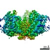
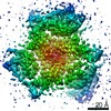
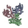
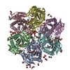
- Assembly
| Deposited unit | 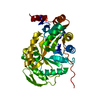
| ||||||||
|---|---|---|---|---|---|---|---|---|---|
| 1 | 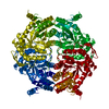
| ||||||||
| 2 | 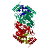
| ||||||||
| Unit cell |
| ||||||||
| Details | This protein exists as monomer |
-Components
| #1: Protein | Mass: 41783.105 Da / Num. of mol.: 1 Source method: isolated from a genetically manipulated source Source: (gene. exp.) References: UniProt: P80645, Oxidoreductases; Acting on paired donors, with incorporation or reduction of molecular oxygen; With reduced flavin or flavoprotein as one donor, and incorporation of one ...References: UniProt: P80645, Oxidoreductases; Acting on paired donors, with incorporation or reduction of molecular oxygen; With reduced flavin or flavoprotein as one donor, and incorporation of one atom of oxygen into the other donor |
|---|---|
| #2: Water | ChemComp-HOH /  Mass: 18.015 Da / Num. of mol.: 144 / Source method: isolated from a natural source / Formula: H2O Mass: 18.015 Da / Num. of mol.: 144 / Source method: isolated from a natural source / Formula: H2O |
-Experimental details
-Experiment
| Experiment | Method: X-RAY DIFFRACTION / Number of used crystals: 1 |
|---|
- Sample preparation
| Crystal | Density Matthews: 2.48 Å3/Da / Density % sol: 50.49 % |
|---|---|
| Crystal grow | Temperature: 298 K / Method: vapor diffusion, hanging drop / pH: 4.6 Details: 0.2M NH4 Acetate, 0.1 M Ma Acetate. 22.5% PEG4000, pH 4.6, VAPOR DIFFUSION, HANGING DROP, temperature 298K |
-Data collection
| Diffraction | Mean temperature: 100 K | ||||||||||||
|---|---|---|---|---|---|---|---|---|---|---|---|---|---|
| Diffraction source | Source: SYNCHROTRON / Site: APS  / Beamline: 19-ID / Wavelength: 0.9793,0.9791,0.9639 / Beamline: 19-ID / Wavelength: 0.9793,0.9791,0.9639 | ||||||||||||
| Detector | Type: SBC-2 / Detector: CCD / Date: Nov 24, 2002 / Details: Si 111 Channel | ||||||||||||
| Radiation | Monochromator: Si 111 Channel / Protocol: MAD / Monochromatic (M) / Laue (L): M / Scattering type: x-ray | ||||||||||||
| Radiation wavelength |
| ||||||||||||
| Reflection | Resolution: 2.18→50 Å / Num. all: 22690 / Num. obs: 22467 / % possible obs: 99 % / Observed criterion σ(F): 2 / Observed criterion σ(I): 2 / Redundancy: 12.89 % / Biso Wilson estimate: 24.7 Å2 / Rmerge(I) obs: 0.072 / Net I/σ(I): 33.87 | ||||||||||||
| Reflection shell | Resolution: 2.18→2.26 Å / Redundancy: 11.9 % / Rmerge(I) obs: 0.56 / Mean I/σ(I) obs: 2.08 / Num. unique all: 2281 / % possible all: 92.3 |
- Processing
| Software |
| ||||||||||||||||||||||||||||||||||||
|---|---|---|---|---|---|---|---|---|---|---|---|---|---|---|---|---|---|---|---|---|---|---|---|---|---|---|---|---|---|---|---|---|---|---|---|---|---|
| Refinement | Method to determine structure: MAD Starting model: none Resolution: 2.2→42.13 Å / Rfactor Rfree error: 0.006 / Isotropic thermal model: RESTRAINED / Cross valid method: THROUGHOUT / σ(F): 0 / σ(I): 0 / Stereochemistry target values: Engh & Huber Details: The number of reflections for refinement is greater than the number of reflections for data collection, because in CNS (hlml taget) refinement, the Friedel's pair was treated as two seperated reflections.
| ||||||||||||||||||||||||||||||||||||
| Solvent computation | Solvent model: FLAT MODEL / Bsol: 54.8496 Å2 / ksol: 0.363014 e/Å3 | ||||||||||||||||||||||||||||||||||||
| Displacement parameters | Biso mean: 44.3 Å2
| ||||||||||||||||||||||||||||||||||||
| Refine analyze | Luzzati coordinate error free: 0.36 Å / Luzzati sigma a free: 0.3 Å | ||||||||||||||||||||||||||||||||||||
| Refinement step | Cycle: LAST / Resolution: 2.2→42.13 Å
| ||||||||||||||||||||||||||||||||||||
| Refine LS restraints |
| ||||||||||||||||||||||||||||||||||||
| LS refinement shell | Resolution: 2.2→2.34 Å / Rfactor Rfree error: 0.021 / Total num. of bins used: 6
| ||||||||||||||||||||||||||||||||||||
| Xplor file |
|
