 Movie
Movie Controller


+ Open data

- Basic information
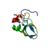
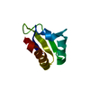
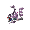
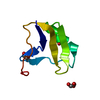
| Entry | Database: PDB / ID: 1nps | ||||||
|---|---|---|---|---|---|---|---|
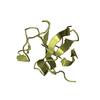
| Title | CRYSTAL STRUCTURE OF N-TERMINAL DOMAIN OF PROTEIN S | ||||||
 Components Components | DEVELOPMENT-SPECIFIC PROTEIN S | ||||||
 Keywords Keywords | SIGNALING PROTEIN / CRYSTALLINE LIKE STRUCTURE | ||||||
| Function / homology |  Function and homology information Function and homology informationsporulation resulting in formation of a cellular spore / metal ion binding Similarity search - Function | ||||||
| Biological species |  Myxococcus xanthus (bacteria) Myxococcus xanthus (bacteria) | ||||||
| Method |  X-RAY DIFFRACTION / MOLECULAR REPLACEMENT / Resolution: 1.8 Å X-RAY DIFFRACTION / MOLECULAR REPLACEMENT / Resolution: 1.8 Å | ||||||
 Authors Authors | Wenk, M. / Baumgartner, R. / Mayer, E.M. / Huber, R. / Holak, T.A. / Jaenicke, R. | ||||||
 Citation Citation | Journal: J.Mol.Biol. / Year: 1999 Title: The domains of protein S from Myxococcus xanthus: structure, stability and interactions. Authors: Wenk, M. / Baumgartner, R. / Holak, T.A. / Huber, R. / Jaenicke, R. / Mayr, E.M. #1: Journal: Structure / Year: 1994Title: NMR-Derived Three-Dimensional Solution Structure of Protein S Complexed with Calcium Authors: Bagby, S. / Harvey, T.S. / Eagle, S.G. / Inouye, S. / Ikura, M. | ||||||
| History |
|
- Structure visualization
| Structure viewer | Molecule: MolmilJmol/JSmol |
|---|
- Downloads & links
-Download
| PDBx/mmCIF format | 1nps.cif.gz | 30.1 KB | Display | PDBx/mmCIF format |
|---|---|---|---|---|
| PDB format | pdb1nps.ent.gz | 19.5 KB | Display | PDB format |
| PDBx/mmJSON format | 1nps.json.gz | Tree view | PDBx/mmJSON format | |
| Others |  Other downloads Other downloads |
-Validation report
| Arichive directory | https://data.pdbj.org/pub/pdb/validation_reports/np/1npsftp://data.pdbj.org/pub/pdb/validation_reports/np/1nps | HTTPS FTP |
|---|
-Related structure data
| Similar structure data |
|---|


-Links
 PDBj
PDBj
- Assembly
| Deposited unit | 
| ||||||||
|---|---|---|---|---|---|---|---|---|---|
| 1 |
| ||||||||
| Unit cell |
|
-Components
| #1: Protein | Mass: 9389.440 Da / Num. of mol.: 1 / Fragment: N-TERMINAL DOMAIN: MOTIFS 1-2 AND LINKER Source method: isolated from a genetically manipulated source Source: (gene. exp.) Myxococcus xanthus (bacteria) / Cellular location: CYTOSOL / Species (production host): Escherichia coli / Cellular location (production host): CYTOPLASM / Production host: | ||
|---|---|---|---|
| #2: Chemical |   Mass: 40.078 Da / Num. of mol.: 2 / Source method: obtained synthetically / Formula: Ca Mass: 40.078 Da / Num. of mol.: 2 / Source method: obtained synthetically / Formula: Ca#3: Water | ChemComp-HOH / |  Mass: 18.015 Da / Num. of mol.: 76 / Source method: isolated from a natural source / Formula: H2O Mass: 18.015 Da / Num. of mol.: 76 / Source method: isolated from a natural source / Formula: H2O |
-Experimental details
-Experiment
| Experiment | Method: X-RAY DIFFRACTION / Number of used crystals: 1 |
|---|
- Sample preparation
| Crystal | Density Matthews: 2.05 Å3/Da / Density % sol: 40.05 % | ||||||||||||||||||||||||
|---|---|---|---|---|---|---|---|---|---|---|---|---|---|---|---|---|---|---|---|---|---|---|---|---|---|
| Crystal grow | pH: 8 / Details: pH 8.0 | ||||||||||||||||||||||||
| Crystal | *PLUS Density % sol: 36 % | ||||||||||||||||||||||||
| Crystal grow | *PLUS Method: vapor diffusion, hanging drop / pH: 7.5 | ||||||||||||||||||||||||
| Components of the solutions | *PLUS
|
-Data collection
| Diffraction | Mean temperature: 287 K |
|---|---|
| Diffraction source | Source: ROTATING ANODE / Type: RIGAKU RU200 / Wavelength: 1.54 |
| Detector | Type: MARRESEARCH / Detector: AREA DETECTOR / Date: Jul 1, 1998 / Details: MIRRORS |
| Radiation | Monochromator: NI FILTER / Protocol: SINGLE WAVELENGTH / Monochromatic (M) / Laue (L): M / Scattering type: x-ray |
| Radiation wavelength | Wavelength: 1.54 Å / Relative weight: 1 |
| Reflection | Resolution: 1.8→20 Å / Num. obs: 7166 / % possible obs: 99 % / Observed criterion σ(I): 2 / Redundancy: 2.3 % / Rmerge(I) obs: 0.08 |
- Processing
| Software |
| |||||||||||||||||||||||||||||||||||||||||||||||||||||||||||||||
|---|---|---|---|---|---|---|---|---|---|---|---|---|---|---|---|---|---|---|---|---|---|---|---|---|---|---|---|---|---|---|---|---|---|---|---|---|---|---|---|---|---|---|---|---|---|---|---|---|---|---|---|---|---|---|---|---|---|---|---|---|---|---|---|---|
| Refinement | Method to determine structure: MOLECULAR REPLACEMENT / Resolution: 1.8→20 Å / Cross valid method: THROUGHOUT / σ(F): 1
| |||||||||||||||||||||||||||||||||||||||||||||||||||||||||||||||
| Refinement step | Cycle: LAST / Resolution: 1.8→20 Å
| |||||||||||||||||||||||||||||||||||||||||||||||||||||||||||||||
| Refine LS restraints |
| |||||||||||||||||||||||||||||||||||||||||||||||||||||||||||||||
| Software | *PLUS Name: REFMAC / Classification: refinement | |||||||||||||||||||||||||||||||||||||||||||||||||||||||||||||||
| Refinement | *PLUS Highest resolution: 1.8 Å / Lowest resolution: 20 Å / σ(F): 1 / % reflection Rfree: 5 % / Rfactor obs: 0.198 / Rfactor Rwork: 0.2 | |||||||||||||||||||||||||||||||||||||||||||||||||||||||||||||||
| Solvent computation | *PLUS | |||||||||||||||||||||||||||||||||||||||||||||||||||||||||||||||
| Displacement parameters | *PLUS | |||||||||||||||||||||||||||||||||||||||||||||||||||||||||||||||
| Refine LS restraints | *PLUS Type: p_angle_d / Dev ideal: 0.9 |
