 Movie
Movie Controller

[English] 日本語
 Yorodumi
- PDB-1njj: Crystal structure determination of T. brucei ornithine decarboxyl... -
+ Open data

- Basic information
| Entry | Database: PDB / ID: 1njj | ||||||
|---|---|---|---|---|---|---|---|
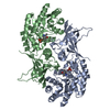
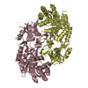
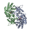
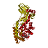
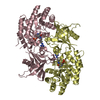
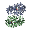
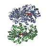
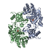
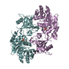
| Title | Crystal structure determination of T. brucei ornithine decarboxylase bound to D-ornithine and to G418 | ||||||
 Components Components | ornithine decarboxylase | ||||||
 Keywords Keywords | LYASE / ornithine decarboxylase / ODC / PLP / pyridoxal 5'-phosphate / D-ornithine / G418 | ||||||
| Function / homology |  Function and homology information Function and homology informationornithine decarboxylase / : / ornithine decarboxylase activity / cytoplasm Similarity search - Function | ||||||
| Biological species |  | ||||||
| Method |  X-RAY DIFFRACTION / MOLECULAR REPLACEMENT / Resolution: 2.45 Å X-RAY DIFFRACTION / MOLECULAR REPLACEMENT / Resolution: 2.45 Å | ||||||
 Authors Authors | Jackson, L.K. / Goldsmith, E.J. / Phillips, M.A. | ||||||
 Citation Citation | Journal: J.Biol.Chem. / Year: 2003 Title: X-ray Structure Determination of Trypanosoma brucei Ornithine Decarboxylase Bound to D-Ornithine and to G418: INSIGHTS INTO SUBSTRATE BINDING AND ODC CONFORMATIONAL FLEXIBILITY. Authors: Jackson, L.K. / Goldsmith, E.J. / Phillips, M.A. | ||||||
| History |
|
- Structure visualization
| Structure viewer | Molecule: MolmilJmol/JSmol |
|---|
- Downloads & links
-Download
| PDBx/mmCIF format | 1njj.cif.gz | 290.5 KB | Display | PDBx/mmCIF format |
|---|---|---|---|---|
| PDB format | pdb1njj.ent.gz | 234.8 KB | Display | PDB format |
| PDBx/mmJSON format | 1njj.json.gz | Tree view | PDBx/mmJSON format | |
| Others |  Other downloads Other downloads |
-Validation report
| Arichive directory | https://data.pdbj.org/pub/pdb/validation_reports/nj/1njjftp://data.pdbj.org/pub/pdb/validation_reports/nj/1njj | HTTPS FTP |
|---|
-Related structure data
| Related structure data |  1f3tS S: Starting model for refinement |
|---|---|
| Similar structure data |


-Links
 PDBj
PDBj- Assembly
| Deposited unit | 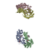
| ||||||||
|---|---|---|---|---|---|---|---|---|---|
| 1 | 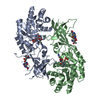
| ||||||||
| 2 | 
| ||||||||
| Unit cell |
|
-Components
| #1: Protein | Mass: 47150.590 Da / Num. of mol.: 4 Source method: isolated from a genetically manipulated source Source: (gene. exp.) Species: Trypanosoma brucei / Strain: gambiense / Plasmid: PODC29 / Species (production host): Escherichia coli / Production host:  #2: Chemical |   Mass: 496.552 Da / Num. of mol.: 3 / Source method: obtained synthetically / Formula: C20H40N4O10 / Comment: antibiotic*YM Mass: 496.552 Da / Num. of mol.: 3 / Source method: obtained synthetically / Formula: C20H40N4O10 / Comment: antibiotic*YM#3: Chemical | ChemComp-ORX /   Mass: 363.303 Da / Num. of mol.: 4 / Source method: obtained synthetically / Formula: C13H22N3O7P Mass: 363.303 Da / Num. of mol.: 4 / Source method: obtained synthetically / Formula: C13H22N3O7P#4: Water | ChemComp-HOH / |  Mass: 18.015 Da / Num. of mol.: 207 / Source method: isolated from a natural source / Formula: H2O Mass: 18.015 Da / Num. of mol.: 207 / Source method: isolated from a natural source / Formula: H2O |
|---|
-Experimental details
-Experiment
| Experiment | Method: X-RAY DIFFRACTION / Number of used crystals: 1 |
|---|
- Sample preparation
| Crystal | Density Matthews: 2.82 Å3/Da / Density % sol: 56.11 % | ||||||||||||||||||||||||||||||||||||||||||||||||||||||||||||||||||||||||||||||||||||||||||||||||||
|---|---|---|---|---|---|---|---|---|---|---|---|---|---|---|---|---|---|---|---|---|---|---|---|---|---|---|---|---|---|---|---|---|---|---|---|---|---|---|---|---|---|---|---|---|---|---|---|---|---|---|---|---|---|---|---|---|---|---|---|---|---|---|---|---|---|---|---|---|---|---|---|---|---|---|---|---|---|---|---|---|---|---|---|---|---|---|---|---|---|---|---|---|---|---|---|---|---|---|---|
| Crystal grow | Temperature: 289 K / Method: vapor diffusion, sitting drop / pH: 7 Details: Peg 3350, Hepes, DTT, NaCl, D-ornithine, G418, butyrolactone, pH 7, VAPOR DIFFUSION, SITTING DROP, temperature 16K | ||||||||||||||||||||||||||||||||||||||||||||||||||||||||||||||||||||||||||||||||||||||||||||||||||
| Crystal grow | *PLUS pH: 7.2 / Method: vapor diffusion | ||||||||||||||||||||||||||||||||||||||||||||||||||||||||||||||||||||||||||||||||||||||||||||||||||
| Components of the solutions | *PLUS
|
-Data collection
| Diffraction | Mean temperature: 98.5 K |
|---|---|
| Diffraction source | Source: ROTATING ANODE / Type: RIGAKU RU200 / Wavelength: 1.5418 |
| Detector | Type: RIGAKU RAXIS IV / Detector: IMAGE PLATE / Date: Apr 1, 2001 |
| Radiation | Protocol: SINGLE WAVELENGTH / Monochromatic (M) / Laue (L): M / Scattering type: x-ray |
| Radiation wavelength | Wavelength: 1.5418 Å / Relative weight: 1 |
| Reflection | Resolution: 2.45→50 Å / Num. obs: 63372 / % possible obs: 96.7 % / Redundancy: 4 % / Biso Wilson estimate: 52 Å2 / Rmerge(I) obs: 0.069 / Net I/σ(I): 21.8 |
| Reflection shell | Resolution: 2.45→2.54 Å / Rmerge(I) obs: 0.62 / Mean I/σ(I) obs: 2.1 / % possible all: 93.9 |
| Reflection | *PLUS Num. measured all: 253681 |
| Reflection shell | *PLUS % possible obs: 93.9 % |
- Processing
| Software |
| |||||||||||||||||||||||||
|---|---|---|---|---|---|---|---|---|---|---|---|---|---|---|---|---|---|---|---|---|---|---|---|---|---|---|
| Refinement | Method to determine structure: MOLECULAR REPLACEMENT Starting model: PDB entry 1f3t with waters, K69, C360 and putrescine removed Resolution: 2.45→35 Å / σ(F): 2 / σ(I): 2
| |||||||||||||||||||||||||
| Displacement parameters | Biso mean: 45.1 Å2 | |||||||||||||||||||||||||
| Refinement step | Cycle: LAST / Resolution: 2.45→35 Å
| |||||||||||||||||||||||||
| LS refinement shell | Resolution: 2.45→2.54 Å
| |||||||||||||||||||||||||
| Refinement | *PLUS Rfactor Rwork: 0.26 | |||||||||||||||||||||||||
| Solvent computation | *PLUS | |||||||||||||||||||||||||
| Displacement parameters | *PLUS | |||||||||||||||||||||||||
| Refine LS restraints | *PLUS
|
