 Movie
Movie Controller

[English] 日本語
 Yorodumi
- PDB-1njg: Nucleotide-free form of an Isolated E. coli Clamp Loader Gamma Subunit -
+ Open data

- Basic information
| Entry | Database: PDB / ID: 1njg | ||||||
|---|---|---|---|---|---|---|---|
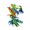
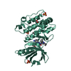
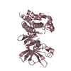
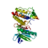
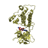
| Title | Nucleotide-free form of an Isolated E. coli Clamp Loader Gamma Subunit | ||||||
 Components Components | DNA polymerase III subunit gamma | ||||||
 Keywords Keywords | TRANSFERASE / Rossman-like fold / AAA+ ATPase domains / sensor 1 / sensor 2 | ||||||
| Function / homology |  Function and homology information Function and homology informationDNA polymerase III, clamp loader complex / DNA clamp loader activity / DNA polymerase III complex / replisome / DNA polymerase processivity factor activity / DNA-templated DNA replication / ribonucleoside triphosphate phosphatase activity / DNA-directed DNA polymerase / DNA-directed DNA polymerase activity / DNA replication ...DNA polymerase III, clamp loader complex / DNA clamp loader activity / DNA polymerase III complex / replisome / DNA polymerase processivity factor activity / DNA-templated DNA replication / ribonucleoside triphosphate phosphatase activity / DNA-directed DNA polymerase / DNA-directed DNA polymerase activity / DNA replication / viral translational frameshifting / ATP hydrolysis activity / DNA binding / ATP binding / metal ion binding / identical protein binding Similarity search - Function | ||||||
| Biological species |  | ||||||
| Method |  X-RAY DIFFRACTION / SYNCHROTRON / MOLECULAR REPLACEMENT / Resolution: 2.2 Å X-RAY DIFFRACTION / SYNCHROTRON / MOLECULAR REPLACEMENT / Resolution: 2.2 Å | ||||||
 Authors Authors | Podobnik, M. / Weitze, T.F. / O'Donnell, M. / Kuriyan, J. | ||||||
 Citation Citation | Journal: Structure / Year: 2003 Title: Nucleotide-Induced Conformational Changes in an Isolated Escherichia coli DNA Polymerase III Clamp Loader Subunit Authors: Podobnik, M. / Weitze, T.F. / O'Donnell, M. / Kuriyan, J. | ||||||
| History |
|
- Structure visualization
| Structure viewer | Molecule: MolmilJmol/JSmol |
|---|
- Downloads & links
-Download
| PDBx/mmCIF format | 1njg.cif.gz | 107.1 KB | Display | PDBx/mmCIF format |
|---|---|---|---|---|
| PDB format | pdb1njg.ent.gz | 82.5 KB | Display | PDB format |
| PDBx/mmJSON format | 1njg.json.gz | Tree view | PDBx/mmJSON format | |
| Others |  Other downloads Other downloads |
-Validation report
| Arichive directory | https://data.pdbj.org/pub/pdb/validation_reports/nj/1njgftp://data.pdbj.org/pub/pdb/validation_reports/nj/1njg | HTTPS FTP |
|---|
-Related structure data
| Related structure data | 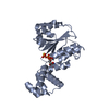 1njfC 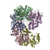 1jr3S C: citing same article ( S: Starting model for refinement |
|---|---|
| Similar structure data |


-Links
 PDBj
PDBj
- Assembly
| Deposited unit | 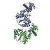
| ||||||||
|---|---|---|---|---|---|---|---|---|---|
| 1 | 
| ||||||||
| 2 | 
| ||||||||
| Unit cell |
| ||||||||
| Details | There are two copies (chains A and B) of the monomeric biological unit in the asymmetric unit. |
-Components
| #1: Protein | Mass: 27499.430 Da / Num. of mol.: 2 / Fragment: N-terminal domains 1 and 2 Source method: isolated from a genetically manipulated source Source: (gene. exp.) #2: Chemical |   Mass: 65.409 Da / Num. of mol.: 2 / Source method: obtained synthetically / Formula: Zn Mass: 65.409 Da / Num. of mol.: 2 / Source method: obtained synthetically / Formula: Zn#3: Chemical |   Mass: 96.063 Da / Num. of mol.: 3 / Source method: obtained synthetically / Formula: SO4 Mass: 96.063 Da / Num. of mol.: 3 / Source method: obtained synthetically / Formula: SO4#4: Water | ChemComp-HOH / |  Mass: 18.015 Da / Num. of mol.: 130 / Source method: isolated from a natural source / Formula: H2O Mass: 18.015 Da / Num. of mol.: 130 / Source method: isolated from a natural source / Formula: H2O |
|---|
-Experimental details
-Experiment
| Experiment | Method: X-RAY DIFFRACTION / Number of used crystals: 1 |
|---|
- Sample preparation
| Crystal | Density Matthews: 2.13 Å3/Da / Density % sol: 41.71 % | ||||||||||||||||||||||||||||||||||||
|---|---|---|---|---|---|---|---|---|---|---|---|---|---|---|---|---|---|---|---|---|---|---|---|---|---|---|---|---|---|---|---|---|---|---|---|---|---|
| Crystal grow | Temperature: 293 K / Method: vapor diffusion, hanging drop / pH: 6 Details: PEG 3350, ammonium sulfate, pH 6.0, VAPOR DIFFUSION, HANGING DROP, temperature 293K | ||||||||||||||||||||||||||||||||||||
| Crystal grow | *PLUS Method: unknown | ||||||||||||||||||||||||||||||||||||
| Components of the solutions | *PLUS
|
-Data collection
| Diffraction | Mean temperature: 100 K |
|---|---|
| Diffraction source | Source: SYNCHROTRON / Site: ALS  / Beamline: 8.3.1 / Wavelength: 1 Å / Beamline: 8.3.1 / Wavelength: 1 Å |
| Detector | Type: ADSC QUANTUM 210 / Detector: CCD / Date: Feb 27, 2002 |
| Radiation | Monochromator: Double-crystal Si(111) / Protocol: SINGLE WAVELENGTH / Monochromatic (M) / Laue (L): M / Scattering type: x-ray |
| Radiation wavelength | Wavelength: 1 Å / Relative weight: 1 |
| Reflection | Resolution: 2.2→100 Å / Num. obs: 21425 / Observed criterion σ(F): -1.5 / Observed criterion σ(I): -3 / Redundancy: 3 % / Biso Wilson estimate: 23.6 Å2 / Rmerge(I) obs: 0.04 / Net I/σ(I): 20.9 |
| Reflection shell | Resolution: 2.2→2.25 Å / Redundancy: 2.8 % / Rmerge(I) obs: 0.338 / Mean I/σ(I) obs: 2.8 / % possible all: 79 |
| Reflection | *PLUS Lowest resolution: 100 Å / Num. obs: 23273 / % possible obs: 92.1 % / Num. measured all: 255606 / Rmerge(I) obs: 0.04 |
| Reflection shell | *PLUS % possible obs: 79 % |
- Processing
| Software |
| ||||||||||||||||||||||||||||||||||||||||||||||||||||||||||||||||||||||||||||||||
|---|---|---|---|---|---|---|---|---|---|---|---|---|---|---|---|---|---|---|---|---|---|---|---|---|---|---|---|---|---|---|---|---|---|---|---|---|---|---|---|---|---|---|---|---|---|---|---|---|---|---|---|---|---|---|---|---|---|---|---|---|---|---|---|---|---|---|---|---|---|---|---|---|---|---|---|---|---|---|---|---|---|
| Refinement | Method to determine structure: MOLECULAR REPLACEMENT Starting model: PDB entry 1JR3 Resolution: 2.2→56.85 Å / Rfactor Rfree error: 0.006 / Data cutoff high absF: 366008.63 / Data cutoff high rms absF: 366008.63 / Data cutoff low absF: 0 / Isotropic thermal model: RESTRAINED / Cross valid method: THROUGHOUT / σ(F): 0 / Stereochemistry target values: Engh & Huber
| ||||||||||||||||||||||||||||||||||||||||||||||||||||||||||||||||||||||||||||||||
| Solvent computation | Solvent model: FLAT MODEL / Bsol: 44.1996 Å2 / ksol: 0.358159 e/Å3 | ||||||||||||||||||||||||||||||||||||||||||||||||||||||||||||||||||||||||||||||||
| Displacement parameters | Biso mean: 47.6 Å2
| ||||||||||||||||||||||||||||||||||||||||||||||||||||||||||||||||||||||||||||||||
| Refine analyze |
| ||||||||||||||||||||||||||||||||||||||||||||||||||||||||||||||||||||||||||||||||
| Refinement step | Cycle: LAST / Resolution: 2.2→56.85 Å
| ||||||||||||||||||||||||||||||||||||||||||||||||||||||||||||||||||||||||||||||||
| Refine LS restraints |
| ||||||||||||||||||||||||||||||||||||||||||||||||||||||||||||||||||||||||||||||||
| LS refinement shell | Resolution: 2.2→2.28 Å / Rfactor Rfree error: 0.025 / Total num. of bins used: 10
| ||||||||||||||||||||||||||||||||||||||||||||||||||||||||||||||||||||||||||||||||
| Xplor file |
| ||||||||||||||||||||||||||||||||||||||||||||||||||||||||||||||||||||||||||||||||
| Refinement | *PLUS % reflection Rfree: 10 % / Rfactor Rfree: 0.265 / Rfactor Rwork: 0.235 | ||||||||||||||||||||||||||||||||||||||||||||||||||||||||||||||||||||||||||||||||
| Solvent computation | *PLUS | ||||||||||||||||||||||||||||||||||||||||||||||||||||||||||||||||||||||||||||||||
| Displacement parameters | *PLUS | ||||||||||||||||||||||||||||||||||||||||||||||||||||||||||||||||||||||||||||||||
| Refine LS restraints | *PLUS
|
