 Movie
Movie Controller

[English] 日本語
 Yorodumi
- PDB-1niv: MANNOSE-SPECIFIC AGGLUTININ (LECTIN) FROM SNOWDROP (GALANTHUS NIV... -
+ Open data

- Basic information
| Entry | Database: PDB / ID: 1niv | |||||||||
|---|---|---|---|---|---|---|---|---|---|---|
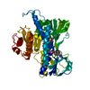
| Title | MANNOSE-SPECIFIC AGGLUTININ (LECTIN) FROM SNOWDROP (GALANTHUS NIVALIS) BULBS IN COMPLEX WITH MANNOSE-ALPHA 1,3-METHYL-D-MANNOSE | |||||||||
 Components Components | AGGLUTININ | |||||||||
 Keywords Keywords | LECTIN / MANNOSE-BINDING | |||||||||
| Function / homology |  Function and homology information Function and homology informationregulation of defense response to virus / D-mannose binding / defense response / response to virus / extracellular region Similarity search - Function | |||||||||
| Biological species |  Galanthus nivalis (common snowdrop) Galanthus nivalis (common snowdrop) | |||||||||
| Method |  X-RAY DIFFRACTION / MOLECULAR REPLACEMENT / Resolution: 3 Å X-RAY DIFFRACTION / MOLECULAR REPLACEMENT / Resolution: 3 Å | |||||||||
 Authors Authors | Wright, C.S. / Hester, G. | |||||||||
 Citation Citation | Journal: J.Mol.Biol. / Year: 1996 Title: The mannose-specific bulb lectin from Galanthus nivalis (snowdrop) binds mono- and dimannosides at distinct sites. Structure analysis of refined complexes at 2.3 A and 3.0 A resolution. Authors: Hester, G. / Wright, C.S. #1: Journal: Nat.Struct.Biol. / Year: 1995Title: Structure of Mannose-Specific Snowdrop (Galanthus Nivalis) Lectin is Representative of a New Plant Lectin Family Authors: Hester, G. / Kaku, H. / Goldstein, I.J. / Wright, C.S. | |||||||||
| History |
|
- Structure visualization
| Structure viewer | Molecule: MolmilJmol/JSmol |
|---|
- Downloads & links
-Download
| PDBx/mmCIF format | 1niv.cif.gz | 52 KB | Display | PDBx/mmCIF format |
|---|---|---|---|---|
| PDB format | pdb1niv.ent.gz | 38.3 KB | Display | PDB format |
| PDBx/mmJSON format | 1niv.json.gz | Tree view | PDBx/mmJSON format | |
| Others |  Other downloads Other downloads |
-Validation report
| Arichive directory | https://data.pdbj.org/pub/pdb/validation_reports/ni/1nivftp://data.pdbj.org/pub/pdb/validation_reports/ni/1niv | HTTPS FTP |
|---|
-Related structure data
| Similar structure data |
|---|


-Links
 PDBj
PDBj
- Assembly
| Deposited unit | 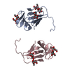
| ||||||||
|---|---|---|---|---|---|---|---|---|---|
| 1 | 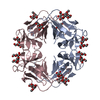
| ||||||||
| Unit cell |
| ||||||||
| Noncrystallographic symmetry (NCS) | NCS oper: (Code: given Matrix: (0.6624, 0.7492, -0.0051), Vector: |
-Components
| #1: Protein | Mass: 12061.348 Da / Num. of mol.: 2 / Source method: isolated from a natural source Details: THE SNOWDROP IS A REPRESENTATIVE OF THE PLANT FAMILY OF AMARYLLIDACEAE Source: (natural) Galanthus nivalis (common snowdrop) / Organ: BULB / References: UniProt: P30617#2: Polysaccharide | Source method: isolated from a genetically manipulated source #3: Sugar |   Type: D-saccharide / Mass: 194.182 Da / Num. of mol.: 3 Type: D-saccharide / Mass: 194.182 Da / Num. of mol.: 3Source method: isolated from a genetically manipulated source Formula: C7H14O6 Has protein modification | Y | |
|---|
-Experimental details
-Experiment
| Experiment | Method: X-RAY DIFFRACTION |
|---|
- Sample preparation
| Crystal | Density Matthews: 4.55 Å3/Da / Density % sol: 68 % | ||||||||||||||||||||
|---|---|---|---|---|---|---|---|---|---|---|---|---|---|---|---|---|---|---|---|---|---|
| Crystal grow | *PLUS pH: 8.5 / Method: unknown | ||||||||||||||||||||
| Components of the solutions | *PLUS
|
-Data collection
| Diffraction source | Wavelength: 1.5418 |
|---|---|
| Detector | Type: RIGAKU / Detector: IMAGE PLATE / Date: Sep 9, 1994 |
| Radiation | Monochromatic (M) / Laue (L): M / Scattering type: x-ray |
| Radiation wavelength | Wavelength: 1.5418 Å / Relative weight: 1 |
| Reflection | Highest resolution: 3 Å / Num. obs: 8280 / % possible obs: 93.1 % / Observed criterion σ(I): 0 / Redundancy: 4.7 % / Rmerge(I) obs: 0.107 / Net I/σ(I): 9 |
| Reflection shell | Resolution: 3→3.11 Å / Redundancy: 2.6 % / Rmerge(I) obs: 0.289 / Mean I/σ(I) obs: 2.9 / % possible all: 86.3 |
| Reflection | *PLUS Num. measured all: 39125 |
| Reflection shell | *PLUS % possible obs: 86.3 % |
- Processing
| Software |
| ||||||||||||||||||||||||||||||||||||||||||||||||||||||||||||
|---|---|---|---|---|---|---|---|---|---|---|---|---|---|---|---|---|---|---|---|---|---|---|---|---|---|---|---|---|---|---|---|---|---|---|---|---|---|---|---|---|---|---|---|---|---|---|---|---|---|---|---|---|---|---|---|---|---|---|---|---|---|
| Refinement | Method to determine structure: MOLECULAR REPLACEMENT Starting model: GNA-ALPHA-MEMAN COMPLEX Resolution: 3→8 Å / σ(F): 0 Details: THE MEAN B-FACTORS OF CHAINS A AND C ARE 22.5 AND 41.7 A**2, RESPECTIVELY. CHAIN C IS VERY FLEXIBLE DUE TO A LACK OF CRYSTAL CONTACTS.
| ||||||||||||||||||||||||||||||||||||||||||||||||||||||||||||
| Displacement parameters | Biso mean: 32.3 Å2 | ||||||||||||||||||||||||||||||||||||||||||||||||||||||||||||
| Refine analyze | Luzzati coordinate error obs: 0.39 Å | ||||||||||||||||||||||||||||||||||||||||||||||||||||||||||||
| Refinement step | Cycle: LAST / Resolution: 3→8 Å
| ||||||||||||||||||||||||||||||||||||||||||||||||||||||||||||
| Refine LS restraints |
| ||||||||||||||||||||||||||||||||||||||||||||||||||||||||||||
| Software | *PLUS Name: X-PLOR / Classification: refinement | ||||||||||||||||||||||||||||||||||||||||||||||||||||||||||||
| Refinement | *PLUS Rfactor all: 0.227 / Rfactor obs: 0.226 | ||||||||||||||||||||||||||||||||||||||||||||||||||||||||||||
| Solvent computation | *PLUS | ||||||||||||||||||||||||||||||||||||||||||||||||||||||||||||
| Displacement parameters | *PLUS | ||||||||||||||||||||||||||||||||||||||||||||||||||||||||||||
| Refine LS restraints | *PLUS
|
