 Movie
Movie Controller

[English] 日本語
 Yorodumi
- PDB-1npl: MANNOSE-SPECIFIC AGGLUTININ (LECTIN) FROM DAFFODIL (NARCISSUS PSE... -
+ Open data

- Basic information
| Entry | Database: PDB / ID: 1npl | |||||||||
|---|---|---|---|---|---|---|---|---|---|---|
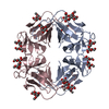
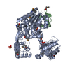
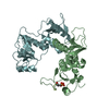
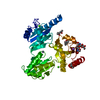
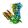
| Title | MANNOSE-SPECIFIC AGGLUTININ (LECTIN) FROM DAFFODIL (NARCISSUS PSEUDONARCISSUS) BULBS IN COMPLEX WITH MANNOSE-ALPHA1,3-MANNOSE | |||||||||
 Components Components | PROTEIN (AGGLUTININ) | |||||||||
 Keywords Keywords | SUGAR BINDING PROTEIN / LECTIN / AGGLUTININ / MANNOBIOSE / MANNOSE-ALPHA1 / 3-MANNOSE / DAFFODIL | |||||||||
| Function / homology |  Function and homology information Function and homology information | |||||||||
| Biological species |  Narcissus pseudonarcissus (daffodil) Narcissus pseudonarcissus (daffodil) | |||||||||
| Method |  X-RAY DIFFRACTION / SYNCHROTRON / MOLECULAR REPLACEMENT / Resolution: 2 Å X-RAY DIFFRACTION / SYNCHROTRON / MOLECULAR REPLACEMENT / Resolution: 2 Å | |||||||||
 Authors Authors | Sauerborn, M.K. / Wright, L.M. / Reynolds, C.D. / Grossmann, J.G. / Rizkallah, P.J. | |||||||||
 Citation Citation | Journal: J.Mol.Biol. / Year: 1999 Title: Insights into carbohydrate recognition by Narcissus pseudonarcissus lectin: the crystal structure at 2 A resolution in complex with alpha1-3 mannobiose. Authors: Sauerborn, M.K. / Wright, L.M. / Reynolds, C.D. / Grossmann, J.G. / Rizkallah, P.J. | |||||||||
| History |
|
- Structure visualization
| Structure viewer | Molecule: MolmilJmol/JSmol |
|---|
- Downloads & links
-Download
| PDBx/mmCIF format | 1npl.cif.gz | 42 KB | Display | PDBx/mmCIF format |
|---|---|---|---|---|
| PDB format | pdb1npl.ent.gz | 29.4 KB | Display | PDB format |
| PDBx/mmJSON format | 1npl.json.gz | Tree view | PDBx/mmJSON format | |
| Others |  Other downloads Other downloads |
-Validation report
| Arichive directory | https://data.pdbj.org/pub/pdb/validation_reports/np/1nplftp://data.pdbj.org/pub/pdb/validation_reports/np/1npl | HTTPS FTP |
|---|
-Related structure data
| Similar structure data |
|---|


-Links
 PDBj
PDBj
- Assembly
| Deposited unit | 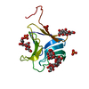
| ||||||||
|---|---|---|---|---|---|---|---|---|---|
| 1 | 
| ||||||||
| 2 | 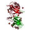
| ||||||||
| Unit cell |
| ||||||||
| Components on special symmetry positions |
|
-Components
| #1: Protein | Mass: 12209.545 Da / Num. of mol.: 1 / Source method: isolated from a natural source / Details: FROM DAFFODIL PLANT FAMILY OF AMARYLLIDACEAE / Source: (natural) Narcissus pseudonarcissus (daffodil) / Organ: BULBS / Strain: DUTCH MASTER / References: GenBank: 289871, UniProt: Q40423*PLUS | ||||||
|---|---|---|---|---|---|---|---|
| #2: Polysaccharide | alpha-D-mannopyranose-(1-3)-alpha-D-mannopyranose / 3alpha-alpha-mannobiose 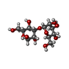  Source method: isolated from a genetically manipulated source Details: oligosaccharide / References: 3alpha-alpha-mannobiose #3: Chemical |   Mass: 94.971 Da / Num. of mol.: 3 / Source method: obtained synthetically / Formula: PO4 Mass: 94.971 Da / Num. of mol.: 3 / Source method: obtained synthetically / Formula: PO4#4: Water | ChemComp-HOH / |  Mass: 18.015 Da / Num. of mol.: 97 / Source method: isolated from a natural source / Formula: H2O Mass: 18.015 Da / Num. of mol.: 97 / Source method: isolated from a natural source / Formula: H2OHas protein modification | Y | |
-Experimental details
-Experiment
| Experiment | Method: X-RAY DIFFRACTION / Number of used crystals: 1 |
|---|
- Sample preparation
| Crystal | Density Matthews: 2.43 Å3/Da / Density % sol: 44 % / Description: ROTATION METHOD | ||||||||||||||||||||||||||||||
|---|---|---|---|---|---|---|---|---|---|---|---|---|---|---|---|---|---|---|---|---|---|---|---|---|---|---|---|---|---|---|---|
| Crystal grow | Temperature: 290 K / Method: vapor diffusion, sitting drop / pH: 6.5 Details: VAPOUR DIFFUSION, SITTING DROP, 10 MG/ML IN PBS CONTAINING UP TO 20 MM MANNOBIOSE, EQUILIBRATED AGAINST 40 - 60% AMMONIUM SULPHATE. 17 DEG. C, 4 - 6 DAYS, pH 6.5, vapor diffusion - sitting drop, temperature 290K | ||||||||||||||||||||||||||||||
| Crystal | *PLUS | ||||||||||||||||||||||||||||||
| Crystal grow | *PLUS Temperature: 17 ℃ | ||||||||||||||||||||||||||||||
| Components of the solutions | *PLUS
|
-Data collection
| Diffraction | Mean temperature: 277 K |
|---|---|
| Diffraction source | Source: SYNCHROTRON / Site: SRS  / Beamline: PX9.6 / Wavelength: 0.87 / Beamline: PX9.6 / Wavelength: 0.87 |
| Detector | Type: MARRESEARCH / Detector: IMAGE PLATE / Date: Sep 19, 1996 / Details: MIRROR, MONOCHROMATOR |
| Radiation | Monochromator: SI (111) / Protocol: SINGLE WAVELENGTH / Monochromatic (M) / Laue (L): M / Scattering type: x-ray |
| Radiation wavelength | Wavelength: 0.87 Å / Relative weight: 1 |
| Reflection | Resolution: 2→60 Å / Num. obs: 9490 / % possible obs: 98.6 % / Redundancy: 2.9 % / Biso Wilson estimate: 20.1 Å2 / Rmerge(I) obs: 0.067 / Rsym value: 0.07 / Net I/σ(I): 7.26 |
| Reflection shell | Resolution: 2→2.12 Å / Redundancy: 2.9 % / Rmerge(I) obs: 0.273 / Mean I/σ(I) obs: 2.2 / Rsym value: 0.276 / % possible all: 98.5 |
| Reflection | *PLUS Num. measured all: 27588 |
| Reflection shell | *PLUS % possible obs: 98.6 % / Num. unique obs: 1322 / Num. measured obs: 4005 |
- Processing
| Software |
| ||||||||||||||||||||||||||||||||||||||||||||||||||||||||||||||||||||||||||||||||||||
|---|---|---|---|---|---|---|---|---|---|---|---|---|---|---|---|---|---|---|---|---|---|---|---|---|---|---|---|---|---|---|---|---|---|---|---|---|---|---|---|---|---|---|---|---|---|---|---|---|---|---|---|---|---|---|---|---|---|---|---|---|---|---|---|---|---|---|---|---|---|---|---|---|---|---|---|---|---|---|---|---|---|---|---|---|---|
| Refinement | Method to determine structure: MOLECULAR REPLACEMENT / Resolution: 2→10 Å / SU B: 4.8 / Cross valid method: THROUGHOUT / σ(F): 0 Details: DISORDERED REGION WERE REFINED IN TWO EQUAL CONFORMATIONS (50% OCCUPANCY EACH). NO ATTEMPT WAS MADE AT REFINING THEIR OCCUPANCIES. THE MODEL WAS CHECKED AGAINST ELECTRON DENSITY.
| ||||||||||||||||||||||||||||||||||||||||||||||||||||||||||||||||||||||||||||||||||||
| Displacement parameters | Biso mean: 27.6 Å2 | ||||||||||||||||||||||||||||||||||||||||||||||||||||||||||||||||||||||||||||||||||||
| Refinement step | Cycle: LAST / Resolution: 2→10 Å
| ||||||||||||||||||||||||||||||||||||||||||||||||||||||||||||||||||||||||||||||||||||
| Refine LS restraints |
| ||||||||||||||||||||||||||||||||||||||||||||||||||||||||||||||||||||||||||||||||||||
| Software | *PLUS Name: REFMAC / Classification: refinement | ||||||||||||||||||||||||||||||||||||||||||||||||||||||||||||||||||||||||||||||||||||
| Refinement | *PLUS Highest resolution: 2 Å / σ(F): 0 / % reflection Rfree: 5 % / Rfactor obs: 0.187 | ||||||||||||||||||||||||||||||||||||||||||||||||||||||||||||||||||||||||||||||||||||
| Solvent computation | *PLUS | ||||||||||||||||||||||||||||||||||||||||||||||||||||||||||||||||||||||||||||||||||||
| Displacement parameters | *PLUS Biso mean: 27.6 Å2 | ||||||||||||||||||||||||||||||||||||||||||||||||||||||||||||||||||||||||||||||||||||
| Refine LS restraints | *PLUS
|
