 Movie
Movie Controller

[English] 日本語
 Yorodumi
- PDB-1nfj: Structure of a Sir2 substrate, alba, reveals a mechanism for deac... -
+ Open data

- Basic information
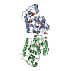
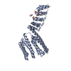
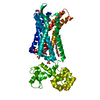
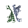
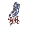
| Entry | Database: PDB / ID: 1nfj | ||||||
|---|---|---|---|---|---|---|---|
| Title | Structure of a Sir2 substrate, alba, reveals a mechanism for deactylation-induced enhancement of DNA-binding | ||||||
 Components Components | conserved hypothetical protein AF1956 | ||||||
 Keywords Keywords | GENE REGULATION / Sir2 / alba / HDAC / transcription | ||||||
| Function / homology |  Function and homology information Function and homology informationchromosome condensation / chromosome / double-stranded DNA binding / RNA binding / cytoplasm Similarity search - Function | ||||||
| Biological species |   Archaeoglobus fulgidus (archaea) Archaeoglobus fulgidus (archaea) | ||||||
| Method |  X-RAY DIFFRACTION / SYNCHROTRON / MAD / Resolution: 2 Å X-RAY DIFFRACTION / SYNCHROTRON / MAD / Resolution: 2 Å | ||||||
 Authors Authors | Zhao, K. / Chai, X. / Marmorstein, R. | ||||||
 Citation Citation | Journal: J.Biol.Chem. / Year: 2003 Title: Structure of a Sir2 substrate, alba, reveals a mechanism for deacetylation-induced enhancement of DNA-binding Authors: Zhao, K. / Chai, X. / Marmorstein, R. | ||||||
| History |
|
- Structure visualization
| Structure viewer | Molecule: MolmilJmol/JSmol |
|---|
- Downloads & links
-Download
| PDBx/mmCIF format | 1nfj.cif.gz | 32.2 KB | Display | PDBx/mmCIF format |
|---|---|---|---|---|
| PDB format | pdb1nfj.ent.gz | 21.9 KB | Display | PDB format |
| PDBx/mmJSON format | 1nfj.json.gz | Tree view | PDBx/mmJSON format | |
| Others |  Other downloads Other downloads |
-Validation report
| Summary document | 1nfj_validation.pdf.gz | 427.8 KB | Display | wwPDB validaton report |
|---|---|---|---|---|
| Full document | 1nfj_full_validation.pdf.gz | 430 KB | Display | |
| Data in XML | 1nfj_validation.xml.gz | 8.3 KB | Display | |
| Data in CIF | 1nfj_validation.cif.gz | 11.2 KB | Display | |
| Arichive directory | https://data.pdbj.org/pub/pdb/validation_reports/nf/1nfjftp://data.pdbj.org/pub/pdb/validation_reports/nf/1nfj | HTTPS FTP |
-Related structure data


-Links
 PDBj
PDBj
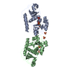
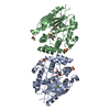
- Assembly
| Deposited unit | 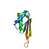
| ||||||||
|---|---|---|---|---|---|---|---|---|---|
| 1 | 
| ||||||||
| Unit cell |
|
-Components
| #1: Protein | Mass: 9908.438 Da / Num. of mol.: 1 Source method: isolated from a genetically manipulated source Source: (gene. exp.) Archaeoglobus fulgidus (archaea) / Plasmid: pGEX 4T-1 / Species (production host): Escherichia coli / Production host:  |
|---|---|
| #2: Water | ChemComp-HOH /  Mass: 18.015 Da / Num. of mol.: 164 / Source method: isolated from a natural source / Formula: H2O Mass: 18.015 Da / Num. of mol.: 164 / Source method: isolated from a natural source / Formula: H2O |
-Experimental details
-Experiment
| Experiment | Method: X-RAY DIFFRACTION / Number of used crystals: 1 |
|---|
- Sample preparation
| Crystal | Density Matthews: 2.16 Å3/Da / Density % sol: 43.01 % | ||||||||||||||||||||||||||||||
|---|---|---|---|---|---|---|---|---|---|---|---|---|---|---|---|---|---|---|---|---|---|---|---|---|---|---|---|---|---|---|---|
| Crystal grow | Temperature: 293 K / Method: vapor diffusion, hanging drop / pH: 8.5 Details: PEG 400, Na-citrate, Tris-HCl, pH 8.5, VAPOR DIFFUSION, HANGING DROP, temperature 293K | ||||||||||||||||||||||||||||||
| Crystal grow | *PLUS Method: vapor diffusion, hanging drop | ||||||||||||||||||||||||||||||
| Components of the solutions | *PLUS
|
-Data collection
| Diffraction | Mean temperature: 100 K |
|---|---|
| Diffraction source | Source: SYNCHROTRON / Site: APS  / Beamline: 19-BM / Wavelength: 0.9795 Å / Beamline: 19-BM / Wavelength: 0.9795 Å |
| Detector | Type: ADSC QUANTUM 4 / Detector: CCD / Date: Aug 10, 2002 |
| Radiation | Monochromator: Ni MIRROR + Ni FILTER / Protocol: SINGLE WAVELENGTH / Monochromatic (M) / Laue (L): M / Scattering type: x-ray |
| Radiation wavelength | Wavelength: 0.9795 Å / Relative weight: 1 |
| Reflection | Resolution: 2→20 Å / Num. obs: 6056 / % possible obs: 99.9 % / Observed criterion σ(F): 2 / Observed criterion σ(I): 0 / Redundancy: 8.6 % / Biso Wilson estimate: 12.8 Å2 / Rmerge(I) obs: 0.058 / Rsym value: 0.056 / Net I/σ(I): 35.6 |
| Reflection shell | Resolution: 2→2.07 Å / Redundancy: 4.5 % / Rmerge(I) obs: 0.179 / Mean I/σ(I) obs: 13.2 / Num. unique all: 580 / Rsym value: 0.173 / % possible all: 100 |
| Reflection shell | *PLUS % possible obs: 100 % |
- Processing
| Software |
| ||||||||||||||||||||||||||||
|---|---|---|---|---|---|---|---|---|---|---|---|---|---|---|---|---|---|---|---|---|---|---|---|---|---|---|---|---|---|
| Refinement | Method to determine structure: MAD / Resolution: 2→20 Å / Cross valid method: THROUGHOUT / σ(F): 0 / Stereochemistry target values: Engh & Huber
| ||||||||||||||||||||||||||||
| Displacement parameters |
| ||||||||||||||||||||||||||||
| Refinement step | Cycle: LAST / Resolution: 2→20 Å
| ||||||||||||||||||||||||||||
| Refine LS restraints |
| ||||||||||||||||||||||||||||
| Xplor file |
| ||||||||||||||||||||||||||||
| Refine LS restraints | *PLUS
|
