 Movie
Movie Controller

[English] 日本語
 Yorodumi
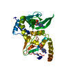
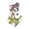
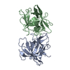
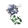
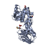
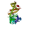
- PDB-1ne6: Crystal structure of Sp-cAMP binding R1a subunit of cAMP-dependen... -
+ Open data

- Basic information
| Entry | Database: PDB / ID: 1ne6 | ||||||
|---|---|---|---|---|---|---|---|
| Title | Crystal structure of Sp-cAMP binding R1a subunit of cAMP-dependent protein kinase | ||||||
 Components Components | cAMP-dependent protein kinase type I-alpha regulatory chain | ||||||
 Keywords Keywords | HYDROLASE / cAMP-dependent protein kinase / R1a subunit / cAMP analog / Sp-cAMP | ||||||
| Function / homology |  Function and homology information Function and homology informationPKA activation in glucagon signalling / DARPP-32 events / CREB1 phosphorylation through the activation of Adenylate Cyclase / GPER1 signaling / Factors involved in megakaryocyte development and platelet production / PKA activation / Hedgehog 'off' state / nucleotide-activated protein kinase complex / cAMP-dependent protein kinase inhibitor activity / cAMP-dependent protein kinase complex ...PKA activation in glucagon signalling / DARPP-32 events / CREB1 phosphorylation through the activation of Adenylate Cyclase / GPER1 signaling / Factors involved in megakaryocyte development and platelet production / PKA activation / Hedgehog 'off' state / nucleotide-activated protein kinase complex / cAMP-dependent protein kinase inhibitor activity / cAMP-dependent protein kinase complex / sarcomere organization / negative regulation of activated T cell proliferation / High laminar flow shear stress activates signaling by PIEZO1 and PECAM1:CDH5:KDR in endothelial cells / Vasopressin regulates renal water homeostasis via Aquaporins / protein kinase A catalytic subunit binding / mesoderm formation / immunological synapse / sperm head-tail coupling apparatus / plasma membrane raft / cardiac muscle cell proliferation / axoneme / cAMP binding / negative regulation of cAMP/PKA signal transduction / multivesicular body / cellular response to glucagon stimulus / neuromuscular junction / positive regulation of insulin secretion / adenylate cyclase-activating G protein-coupled receptor signaling pathway / protein domain specific binding / negative regulation of gene expression / ubiquitin protein ligase binding / centrosome / glutamatergic synapse / identical protein binding / plasma membrane / cytoplasm / cytosol Similarity search - Function | ||||||
| Biological species |  | ||||||
| Method |  X-RAY DIFFRACTION / SYNCHROTRON / MOLECULAR REPLACEMENT / Resolution: 2.3 Å X-RAY DIFFRACTION / SYNCHROTRON / MOLECULAR REPLACEMENT / Resolution: 2.3 Å | ||||||
 Authors Authors | Wu, J. / Jones, J.M. / Xuong, N.H. / Taylor, S.S. | ||||||
 Citation Citation | Journal: Biochemistry / Year: 2004 Title: Crystal Structures of RIalpha Subunit of Cyclic Adenosine 5'-Monophosphate (cAMP)-Dependent Protein Kinase Complexed with (R(p))-Adenosine 3',5'-Cyclic Monophosphothioate and (S(p))-Adenosine ...Title: Crystal Structures of RIalpha Subunit of Cyclic Adenosine 5'-Monophosphate (cAMP)-Dependent Protein Kinase Complexed with (R(p))-Adenosine 3',5'-Cyclic Monophosphothioate and (S(p))-Adenosine 3',5'-Cyclic Monophosphothioate, the Phosphothioate Analogues of cAMP. Authors: Wu, J. / Jones, J.M. / Xuong, N.H. / Eyck, L.F. / Taylor, S.S. | ||||||
| History |
|
- Structure visualization
| Structure viewer | Molecule: MolmilJmol/JSmol |
|---|
- Downloads & links
-Download
| PDBx/mmCIF format | 1ne6.cif.gz | 70.2 KB | Display | PDBx/mmCIF format |
|---|---|---|---|---|
| PDB format | pdb1ne6.ent.gz | 51.3 KB | Display | PDB format |
| PDBx/mmJSON format | 1ne6.json.gz | Tree view | PDBx/mmJSON format | |
| Others |  Other downloads Other downloads |
-Validation report
| Arichive directory | https://data.pdbj.org/pub/pdb/validation_reports/ne/1ne6ftp://data.pdbj.org/pub/pdb/validation_reports/ne/1ne6 | HTTPS FTP |
|---|
-Related structure data
| Related structure data |  1ne4C  1rgsS S: Starting model for refinement C: citing same article ( |
|---|---|
| Similar structure data |


-Links
 PDBj
PDBj


- Assembly
| Deposited unit | 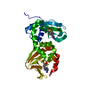
| ||||||||
|---|---|---|---|---|---|---|---|---|---|
| 1 |
| ||||||||
| Unit cell |
|
-Components
| #1: Protein | Mass: 31880.104 Da / Num. of mol.: 1 / Fragment: 1-91 deletion mutant Source method: isolated from a genetically manipulated source Source: (gene. exp.)  | ||
|---|---|---|---|
| #2: Chemical | 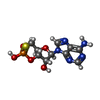  Mass: 345.272 Da / Num. of mol.: 2 / Source method: obtained synthetically / Formula: C10H12N5O5PS Mass: 345.272 Da / Num. of mol.: 2 / Source method: obtained synthetically / Formula: C10H12N5O5PS#3: Water | ChemComp-HOH / |  Mass: 18.015 Da / Num. of mol.: 102 / Source method: isolated from a natural source / Formula: H2O Mass: 18.015 Da / Num. of mol.: 102 / Source method: isolated from a natural source / Formula: H2O |
-Experimental details
-Experiment
| Experiment | Method: X-RAY DIFFRACTION / Number of used crystals: 1 |
|---|
- Sample preparation
| Crystal | Density Matthews: 3.14 Å3/Da / Density % sol: 60.49 % | ||||||||||||||||||||||||||||||||||||||||||
|---|---|---|---|---|---|---|---|---|---|---|---|---|---|---|---|---|---|---|---|---|---|---|---|---|---|---|---|---|---|---|---|---|---|---|---|---|---|---|---|---|---|---|---|
| Crystal grow | Temperature: 295.5 K / Method: vapor diffusion, hanging drop / pH: 5.5 Details: amino sulfate, glycerol, DTT, pH 5.5, VAPOR DIFFUSION, HANGING DROP, temperature 295.5K | ||||||||||||||||||||||||||||||||||||||||||
| Crystal grow | *PLUS Temperature: 22.5 ℃ / Method: vapor diffusion, hanging drop | ||||||||||||||||||||||||||||||||||||||||||
| Components of the solutions | *PLUS
|
-Data collection
| Diffraction | Mean temperature: 200 K |
|---|---|
| Diffraction source | Source: SYNCHROTRON / Site: SSRL  / Beamline: BL7-1 / Beamline: BL7-1 |
| Detector | Type: MARRESEARCH / Detector: IMAGE PLATE / Date: Jun 5, 2001 |
| Radiation | Monochromator: graphite / Protocol: SINGLE WAVELENGTH / Monochromatic (M) / Laue (L): M / Scattering type: x-ray |
| Radiation wavelength | Relative weight: 1 |
| Reflection | Resolution: 2.3→50 Å / Num. all: 18621 / Num. obs: 17939 / % possible obs: 99.5 % / Observed criterion σ(F): 2 / Biso Wilson estimate: 35.4 Å2 / Rsym value: 0.045 / Net I/σ(I): 63.5 |
| Reflection shell | Resolution: 2.3→2.38 Å / Mean I/σ(I) obs: 6.6 / Num. unique all: 1585 / Rsym value: 0.451 / % possible all: 97.9 |
| Reflection | *PLUS Highest resolution: 2.3 Å / Lowest resolution: 50 Å / Num. obs: 18563 / Rmerge(I) obs: 0.045 |
| Reflection shell | *PLUS % possible obs: 97.9 % / Rmerge(I) obs: 0.451 |
- Processing
| Software |
| |||||||||||||||||||||||||
|---|---|---|---|---|---|---|---|---|---|---|---|---|---|---|---|---|---|---|---|---|---|---|---|---|---|---|
| Refinement | Method to determine structure: MOLECULAR REPLACEMENT Starting model: PDB entry 1RGS Resolution: 2.3→50 Å / Isotropic thermal model: isotropic / Cross valid method: THROUGHOUT / σ(F): 2 / Stereochemistry target values: Engh & Huber
| |||||||||||||||||||||||||
| Displacement parameters | Biso mean: 59.1 Å2
| |||||||||||||||||||||||||
| Refine analyze | Luzzati coordinate error obs: 0.38 Å / Luzzati d res low obs: 5 Å / Luzzati sigma a obs: 0.39 Å | |||||||||||||||||||||||||
| Refinement step | Cycle: LAST / Resolution: 2.3→50 Å
| |||||||||||||||||||||||||
| Refine LS restraints |
| |||||||||||||||||||||||||
| LS refinement shell | Resolution: 2.3→2.38 Å / Rfactor Rfree error: 0.04
| |||||||||||||||||||||||||
| Refinement | *PLUS Highest resolution: 2.3 Å / Lowest resolution: 50 Å | |||||||||||||||||||||||||
| Solvent computation | *PLUS | |||||||||||||||||||||||||
| Displacement parameters | *PLUS |
