 Movie
Movie Controller

[English] 日本語
 Yorodumi
- PDB-1n8j: Crystal Structure of AhpC with Active Site Cysteine mutated to Se... -
+ Open data

- Basic information
| Entry | Database: PDB / ID: 1n8j | ||||||
|---|---|---|---|---|---|---|---|
| Title | Crystal Structure of AhpC with Active Site Cysteine mutated to Serine (C46S) | ||||||
 Components Components | Alkyl hydroperoxide reductase C22 protein | ||||||
 Keywords Keywords | OXIDOREDUCTASE / AhpC / peroxiredoxin / decamer / antioxidant / peroxidase / alkylhydroperoxide reductase / AhpF | ||||||
| Function / homology |  Function and homology information Function and homology informationNADH-dependent peroxiredoxin / NADH-dependent peroxiredoxin activity / thioredoxin peroxidase activity / cell redox homeostasis / hydrogen peroxide catabolic process / response to oxidative stress / cytosol Similarity search - Function | ||||||
| Biological species |  Salmonella typhimurium (bacteria) Salmonella typhimurium (bacteria) | ||||||
| Method |  X-RAY DIFFRACTION / SYNCHROTRON / MOLECULAR REPLACEMENT / Resolution: 2.17 Å X-RAY DIFFRACTION / SYNCHROTRON / MOLECULAR REPLACEMENT / Resolution: 2.17 Å | ||||||
 Authors Authors | Wood, Z.A. / Poole, L.B. / Karplus, P.A. | ||||||
 Citation Citation | Journal: Science / Year: 2003 Title: Peroxiredoxin Evolution and the Regulation of Hydrogen Peroxide Signaling Authors: Wood, Z.A. / Poole, L.B. / Karplus, P.A. | ||||||
| History |
|
- Structure visualization
| Structure viewer | Molecule: MolmilJmol/JSmol |
|---|
- Downloads & links
-Download
| PDBx/mmCIF format | 1n8j.cif.gz | 736.2 KB | Display | PDBx/mmCIF format |
|---|---|---|---|---|
| PDB format | pdb1n8j.ent.gz | 613.2 KB | Display | PDB format |
| PDBx/mmJSON format | 1n8j.json.gz | Tree view | PDBx/mmJSON format | |
| Others |  Other downloads Other downloads |
-Validation report
| Arichive directory | https://data.pdbj.org/pub/pdb/validation_reports/n8/1n8jftp://data.pdbj.org/pub/pdb/validation_reports/n8/1n8j | HTTPS FTP |
|---|
-Related structure data
| Related structure data |  1kyg S: Starting model for refinement |
|---|---|
| Similar structure data |


-Links
 PDBj
PDBj- Assembly
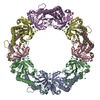
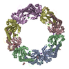
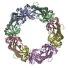
| Deposited unit | 
| ||||||||
|---|---|---|---|---|---|---|---|---|---|
| 1 | 
| ||||||||
| 2 | 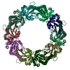
| ||||||||
| Unit cell |
| ||||||||
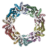
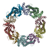
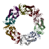
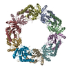
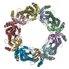
| Details | The biological assembly is a decamer made up of chains A,B,C,D,E,F,G,H,I,J or K,L,M,N,O,P,Q,R,S,T |
-Components
| #1: Protein | Mass: 20625.123 Da / Num. of mol.: 20 / Mutation: C46S Source method: isolated from a genetically manipulated source Source: (gene. exp.) Salmonella typhimurium (bacteria) / Gene: AhpC / Production host: #2: Water | ChemComp-HOH / |  Mass: 18.015 Da / Num. of mol.: 1590 / Source method: isolated from a natural source / Formula: H2O Mass: 18.015 Da / Num. of mol.: 1590 / Source method: isolated from a natural source / Formula: H2O |
|---|
-Experimental details
-Experiment
| Experiment | Method: X-RAY DIFFRACTION / Number of used crystals: 1 |
|---|
- Sample preparation
| Crystal | Density Matthews: 2.67 Å3/Da / Density % sol: 53.6 % | ||||||||||||||||||||||||||||||
|---|---|---|---|---|---|---|---|---|---|---|---|---|---|---|---|---|---|---|---|---|---|---|---|---|---|---|---|---|---|---|---|
| Crystal grow | Temperature: 298 K / Method: vapor diffusion, hanging drop / pH: 4.5 Details: 4% PEG 4000, 10% isopropyl alcohol, 0.1M Na acetate, pH 4.5, VAPOR DIFFUSION, HANGING DROP, temperature 298K | ||||||||||||||||||||||||||||||
| Crystal grow | *PLUS | ||||||||||||||||||||||||||||||
| Components of the solutions | *PLUS
|
-Data collection
| Diffraction | Mean temperature: 100 K |
|---|---|
| Diffraction source | Source: SYNCHROTRON / Site: ALS  / Beamline: 5.0.2 / Wavelength: 1.1 Å / Beamline: 5.0.2 / Wavelength: 1.1 Å |
| Detector | Type: ADSC QUANTUM 4 / Detector: CCD / Date: Jan 19, 2001 / Details: double crystal |
| Radiation | Monochromator: double crystal / Protocol: SINGLE WAVELENGTH / Monochromatic (M) / Laue (L): M / Scattering type: x-ray |
| Radiation wavelength | Wavelength: 1.1 Å / Relative weight: 1 |
| Reflection | Resolution: 2.17→19.99 Å / Num. all: 210095 / Num. obs: 210095 / % possible obs: 89.3 % / Observed criterion σ(F): 0 / Observed criterion σ(I): 0 / Rsym value: 0.066 / Net I/σ(I): 12.1 |
| Reflection shell | Resolution: 2.17→2.31 Å / % possible all: 74.8 |
| Reflection | *PLUS Num. obs: 204205 / % possible obs: 90.4 % / Rmerge(I) obs: 0.074 / Num. measured all: 836640 |
| Reflection shell | *PLUS % possible obs: 82 % / Rmerge(I) obs: 0.47 / Mean I/σ(I) obs: 3.4 |
- Processing
| Software |
| ||||||||||||||||||||||||||||||||||||
|---|---|---|---|---|---|---|---|---|---|---|---|---|---|---|---|---|---|---|---|---|---|---|---|---|---|---|---|---|---|---|---|---|---|---|---|---|---|
| Refinement | Method to determine structure: MOLECULAR REPLACEMENT Starting model: 1KYG 1kyg Resolution: 2.17→19.99 Å / Rfactor Rfree error: 0.002 / Isotropic thermal model: RESTRAINED / Cross valid method: THROUGHOUT / σ(F): 0 / Stereochemistry target values: Engh & Huber
| ||||||||||||||||||||||||||||||||||||
| Solvent computation | Solvent model: FLAT MODEL / Bsol: 43.3883 Å2 / ksol: 0.333622 e/Å3 | ||||||||||||||||||||||||||||||||||||
| Displacement parameters | Biso mean: 47.2 Å2
| ||||||||||||||||||||||||||||||||||||
| Refine analyze |
| ||||||||||||||||||||||||||||||||||||
| Refinement step | Cycle: LAST / Resolution: 2.17→19.99 Å
| ||||||||||||||||||||||||||||||||||||
| Refine LS restraints |
| ||||||||||||||||||||||||||||||||||||
| LS refinement shell | Resolution: 2.17→2.31 Å / Rfactor Rfree error: 0.01 / Total num. of bins used: 6
| ||||||||||||||||||||||||||||||||||||
| Xplor file |
| ||||||||||||||||||||||||||||||||||||
| Refinement | *PLUS Highest resolution: 2.6 Å / Rfactor Rfree: 0.253 / Rfactor Rwork: 0.187 / Lowest resolution: 50 Å | ||||||||||||||||||||||||||||||||||||
| Solvent computation | *PLUS | ||||||||||||||||||||||||||||||||||||
| Displacement parameters | *PLUS | ||||||||||||||||||||||||||||||||||||
| Refine LS restraints | *PLUS
| ||||||||||||||||||||||||||||||||||||
| LS refinement shell | *PLUS Lowest resolution: 2.28 Å / Rfactor Rfree: 0.36 / Rfactor Rwork: 0.335 |
