 Movie
Movie Controller

[English] 日本語
 Yorodumi
- PDB-1n5l: CRYSTAL STRUCTURE OF MYCOBACTERIUM TUBERCULOSIS THYMIDYLATE KINAS... -
+ Open data

- Basic information
| Entry | Database: PDB / ID: 1n5l | ||||||
|---|---|---|---|---|---|---|---|
| Title | CRYSTAL STRUCTURE OF MYCOBACTERIUM TUBERCULOSIS THYMIDYLATE KINASE CRYSTALLIZED IN SODIUM MALONATE, AFTER CATALYSIS IN THE CRYSTAL (2.3 A RESOLUTION) | ||||||
 Components Components | THYMIDYLATE KINASE | ||||||
 Keywords Keywords | TRANSFERASE / TRANSFERASE (ATP:TMP PHOSPHOTRANSFERASE) / KINASE | ||||||
| Function / homology |  Function and homology information Function and homology informationTMP metabolic process / dTMP kinase / dUDP biosynthetic process / dTDP biosynthetic process / dTMP kinase activity / dTTP biosynthetic process / GTP binding / magnesium ion binding / protein homodimerization activity / ATP binding ...TMP metabolic process / dTMP kinase / dUDP biosynthetic process / dTDP biosynthetic process / dTMP kinase activity / dTTP biosynthetic process / GTP binding / magnesium ion binding / protein homodimerization activity / ATP binding / metal ion binding / cytosol / cytoplasm Similarity search - Function | ||||||
| Biological species |   Mycobacterium tuberculosis (bacteria) Mycobacterium tuberculosis (bacteria) | ||||||
| Method |  X-RAY DIFFRACTION / SYNCHROTRON / OTHER / Resolution: 2.3 Å X-RAY DIFFRACTION / SYNCHROTRON / OTHER / Resolution: 2.3 Å | ||||||
 Authors Authors | Fioravanti, E. / Haouz, A. / Ursby, T. / Munier-Lehmann, H. / Delarue, M. / Bourgeois, D. | ||||||
 Citation Citation | Journal: J.Mol.Biol. / Year: 2003 Title: Mycobacterium tuberculosis Thymidylate Kinase: Structural Studies of Intermediates along the Reaction Pathway Authors: Fioravanti, E. / Haouz, A. / Ursby, T. / Munier-Lehmann, H. / Delarue, M. / Bourgeois, D. #1: Journal: Acta Crystallogr.,Sect.D / Year: 2002Title: Cryo-Photolysis of Caged Compounds: A Technique for Trapping Intermediate States in Protein Crystals Authors: Ursby, T. / Weik, M. / Fioravanti, E. / Delarue, M. / Goeldner, M. / Bourgeois, D. #2: Journal: J.Mol.Biol. / Year: 2001Title: X-Ray Structure of Tmp Kinase from Mycobacterium Tuberculosis Complexed with Tmp at 1.95 A Resolution Authors: Li De La Sierra, I. / Munier-Lehmann, H. / Gilles, A.M. / Barzu, O. / Delarue, M. #3: Journal: Acta Crystallogr.,Sect.D / Year: 2000Title: Crystallization and Preliminary X-Ray Analysis of the Thymidylate Kinase from Mycobacterium Tuberculosis Authors: Li DE La Sierra, I. / Munier-Lehmann, H. / Gilles, A.M. / Barzu, O. / Delarue, M. | ||||||
| History |
| ||||||
| Remark 600 | HETEROGEN HOH molecules 140 and 141 were both named 140 in the paper because they occupy the same ...HETEROGEN HOH molecules 140 and 141 were both named 140 in the paper because they occupy the same site in the 2 monomers (A and B), in order to make an easier comparison of the active sites. |
- Structure visualization
| Structure viewer | Molecule: MolmilJmol/JSmol |
|---|
- Downloads & links
-Download
| PDBx/mmCIF format | 1n5l.cif.gz | 93 KB | Display | PDBx/mmCIF format |
|---|---|---|---|---|
| PDB format | pdb1n5l.ent.gz | 70 KB | Display | PDB format |
| PDBx/mmJSON format | 1n5l.json.gz | Tree view | PDBx/mmJSON format | |
| Others |  Other downloads Other downloads |
-Validation report
| Arichive directory | https://data.pdbj.org/pub/pdb/validation_reports/n5/1n5lftp://data.pdbj.org/pub/pdb/validation_reports/n5/1n5l | HTTPS FTP |
|---|
-Related structure data
| Related structure data |  1n5iC 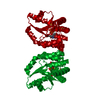 1n5jC 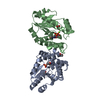 1n5kSC S: Starting model for refinement C: citing same article ( |
|---|---|
| Similar structure data |


-Links
 PDBj
PDBj- Assembly
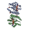
| Deposited unit | 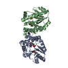
| ||||||||
|---|---|---|---|---|---|---|---|---|---|
| 1 |
| ||||||||
| Unit cell |
| ||||||||
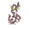
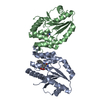
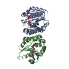
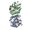
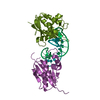
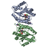
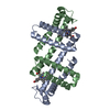
| Details | The asymmetric unit contains the functional dimer. |
-Components
-Protein , 1 types, 2 molecules AB
| #1: Protein | Mass: 22662.525 Da / Num. of mol.: 2 Source method: isolated from a genetically manipulated source Source: (gene. exp.) Mycobacterium tuberculosis (bacteria) / Plasmid: PET22B / Production host: References: UniProt: O05891, UniProt: P9WKE1*PLUS, dTMP kinase |
|---|
-Non-polymers , 6 types, 211 molecules 
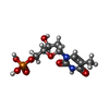


| #2: Chemical |  Mass: 59.044 Da / Num. of mol.: 2 / Source method: obtained synthetically / Formula: C2H3O2 Mass: 59.044 Da / Num. of mol.: 2 / Source method: obtained synthetically / Formula: C2H3O2#3: Chemical | ChemComp-TMP / |  Mass: 322.208 Da / Num. of mol.: 1 / Source method: obtained synthetically / Formula: C10H15N2O8P Mass: 322.208 Da / Num. of mol.: 1 / Source method: obtained synthetically / Formula: C10H15N2O8P#4: Chemical | ChemComp-MG / |  Mass: 24.305 Da / Num. of mol.: 1 / Source method: obtained synthetically / Formula: Mg Mass: 24.305 Da / Num. of mol.: 1 / Source method: obtained synthetically / Formula: Mg#5: Chemical | ChemComp-TYD / |  Mass: 402.188 Da / Num. of mol.: 1 / Source method: obtained synthetically / Formula: C10H16N2O11P2 Mass: 402.188 Da / Num. of mol.: 1 / Source method: obtained synthetically / Formula: C10H16N2O11P2#6: Chemical | ChemComp-DPO / |  Mass: 173.943 Da / Num. of mol.: 1 / Source method: obtained synthetically / Formula: O7P2 Mass: 173.943 Da / Num. of mol.: 1 / Source method: obtained synthetically / Formula: O7P2#7: Water | ChemComp-HOH / | Mass: 18.015 Da / Num. of mol.: 205 / Source method: isolated from a natural source / Formula: H2O |
|---|
-Experimental details
-Experiment
| Experiment | Method: X-RAY DIFFRACTION / Number of used crystals: 1 |
|---|
- Sample preparation
| Crystal | Density Matthews: 2.64 Å3/Da / Density % sol: 53.12 % |
|---|---|
| Crystal grow | Temperature: 293 K Method: vapor diffusion, hanging drop, streak seeding, soaking pH: 6 Details: 1.4 M sodium malonate, 2% PEG 2000, 0.1 M MES pH 6.0, 2 mM b-mercaptoethanol, 25 mM magnesium acetate, soaking in 30 mM ATP, VAPOR DIFFUSION, HANGING DROP, STREAK SEEDING, SOAKING |
-Data collection
| Diffraction | Mean temperature: 100 K |
|---|---|
| Diffraction source | Source: SYNCHROTRON / Site: ESRF  / Beamline: ID29 / Wavelength: 0.936 Å / Beamline: ID29 / Wavelength: 0.936 Å |
| Detector | Type: ADSC QUANTUM 4 / Detector: CCD / Date: Apr 7, 2001 |
| Radiation | Monochromator: silicon / Protocol: SINGLE WAVELENGTH / Monochromatic (M) / Laue (L): M / Scattering type: x-ray |
| Radiation wavelength | Wavelength: 0.936 Å / Relative weight: 1 |
| Reflection | Resolution: 2.3→19.76 Å / Num. all: 21648 / Num. obs: 21648 / % possible obs: 99.4 % / Observed criterion σ(F): 0 / Observed criterion σ(I): -3 / Redundancy: 6.9 % / Biso Wilson estimate: 41 Å2 / Rsym value: 0.107 / Net I/σ(I): 4.6 |
| Reflection shell | Resolution: 2.3→2.42 Å / Redundancy: 7.2 % / Mean I/σ(I) obs: 1.9 / Num. unique all: 2977 / Rsym value: 0.381 / % possible all: 100 |
- Processing
| Software |
| ||||||||||||||||||||||||||||||||||||
|---|---|---|---|---|---|---|---|---|---|---|---|---|---|---|---|---|---|---|---|---|---|---|---|---|---|---|---|---|---|---|---|---|---|---|---|---|---|
| Refinement | Method to determine structure: OTHER Starting model: PDB ENTRY 1N5K Resolution: 2.3→19.76 Å / Rfactor Rfree error: 0.009 / Data cutoff high absF: 1848944.31 / Data cutoff high rms absF: 1848944.31 / Data cutoff low absF: 0 / Isotropic thermal model: RESTRAINED / Cross valid method: THROUGHOUT / σ(F): 0 / Stereochemistry target values: Engh & Huber Details: Monomer A contains the apo-structure. Monomer B contains the TDP-Mg++-ADP complex.
| ||||||||||||||||||||||||||||||||||||
| Solvent computation | Solvent model: FLAT MODEL / Bsol: 63.1841 Å2 / ksol: 0.34923 e/Å3 | ||||||||||||||||||||||||||||||||||||
| Displacement parameters | Biso mean: 35.9 Å2
| ||||||||||||||||||||||||||||||||||||
| Refine analyze |
| ||||||||||||||||||||||||||||||||||||
| Refinement step | Cycle: LAST / Resolution: 2.3→19.76 Å
| ||||||||||||||||||||||||||||||||||||
| Refine LS restraints |
| ||||||||||||||||||||||||||||||||||||
| LS refinement shell | Resolution: 2.3→2.44 Å / Rfactor Rfree error: 0.024 / Total num. of bins used: 6
| ||||||||||||||||||||||||||||||||||||
| Xplor file |
|
