 Movie
Movie Controller

[English] 日本語
 Yorodumi
- PDB-1n48: Y-family DNA polymerase Dpo4 in complex with DNA containing abasi... -
+ Open data

- Basic information
| Entry | Database: PDB / ID: 1n48 | ||||||
|---|---|---|---|---|---|---|---|
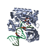
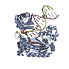
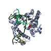
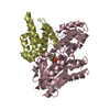
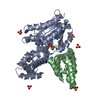
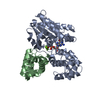
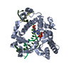
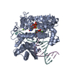
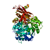
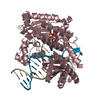
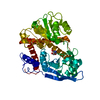
| Title | Y-family DNA polymerase Dpo4 in complex with DNA containing abasic lesion | ||||||
 Components Components |
| ||||||
 Keywords Keywords | TRANSFERASE/DNA / Y-family / DNA polymerase / DNA lesion bypass / protein-DNA complex / TRANSFERASE-DNA COMPLEX | ||||||
| Function / homology |  Function and homology information Function and homology informationerror-prone translesion synthesis / DNA-templated DNA replication / DNA-directed DNA polymerase / damaged DNA binding / DNA-directed DNA polymerase activity / DNA repair / magnesium ion binding / cytoplasm Similarity search - Function | ||||||
| Biological species |   Sulfolobus solfataricus (archaea) Sulfolobus solfataricus (archaea) | ||||||
| Method |  X-RAY DIFFRACTION / MOLECULAR REPLACEMENT / Resolution: 2.2 Å X-RAY DIFFRACTION / MOLECULAR REPLACEMENT / Resolution: 2.2 Å | ||||||
 Authors Authors | Ling, H. / Boudsocq, F. / Woodgate, R. / Yang, W. | ||||||
 Citation Citation | Journal: To be Published Title: A Y-family polymerase complexed with abasic lesions: catching DNA with a loaded nucleoside triphosphate Authors: Ling, H. / Boudsocq, F. / Woodgate, R. / Yang, W. #1: Journal: Cell(Cambridge,Mass.) / Year: 2001Title: Crystal structure of a Y-family DNA polymerase in action: a mechanism for error-prone and lesion-bypass replication. Authors: Ling, H. / Boudsocq, F. / Woodgate, R. / Yang, W. | ||||||
| History |
|
- Structure visualization
| Structure viewer | Molecule: MolmilJmol/JSmol |
|---|
- Downloads & links
-Download
| PDBx/mmCIF format | 1n48.cif.gz | 113.8 KB | Display | PDBx/mmCIF format |
|---|---|---|---|---|
| PDB format | pdb1n48.ent.gz | 81.6 KB | Display | PDB format |
| PDBx/mmJSON format | 1n48.json.gz | Tree view | PDBx/mmJSON format | |
| Others |  Other downloads Other downloads |
-Validation report
| Arichive directory | https://data.pdbj.org/pub/pdb/validation_reports/n4/1n48ftp://data.pdbj.org/pub/pdb/validation_reports/n4/1n48 | HTTPS FTP |
|---|
-Related structure data
| Related structure data | 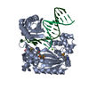 1jx4S S: Starting model for refinement |
|---|---|
| Similar structure data |

-Links
 PDBj
PDBj


- Assembly
| Deposited unit | 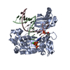
| ||||||||
|---|---|---|---|---|---|---|---|---|---|
| 1 |
| ||||||||
| Unit cell |
|
-Components
-DNA chain , 2 types, 2 molecules BC
| #1: DNA chain | Mass: 4105.685 Da / Num. of mol.: 1 / Source method: obtained synthetically / Details: Template |
|---|---|
| #2: DNA chain | Mass: 4620.983 Da / Num. of mol.: 1 / Source method: obtained synthetically / Details: primer |
-Protein , 1 types, 1 molecules A
| #3: Protein | Mass: 40257.879 Da / Num. of mol.: 1 Source method: isolated from a genetically manipulated source Source: (gene. exp.) Sulfolobus solfataricus (archaea) / Strain: P2 / Gene: dpo4 / Plasmid: pet22bB / Species (production host): Escherichia coli / Production host:  |
|---|
-Non-polymers , 3 types, 393 molecules 


| #4: Chemical |  Mass: 40.078 Da / Num. of mol.: 3 / Source method: obtained synthetically / Formula: Ca Mass: 40.078 Da / Num. of mol.: 3 / Source method: obtained synthetically / Formula: Ca#5: Chemical | ChemComp-ATP / |  Mass: 507.181 Da / Num. of mol.: 1 / Source method: obtained synthetically / Formula: C10H16N5O13P3 / Comment: ATP, energy-carrying molecule*YM Mass: 507.181 Da / Num. of mol.: 1 / Source method: obtained synthetically / Formula: C10H16N5O13P3 / Comment: ATP, energy-carrying molecule*YM#6: Water | ChemComp-HOH / | Mass: 18.015 Da / Num. of mol.: 389 / Source method: isolated from a natural source / Formula: H2O |
|---|
-Experimental details
-Experiment
| Experiment | Method: X-RAY DIFFRACTION / Number of used crystals: 1 |
|---|
- Sample preparation
| Crystal | Density Matthews: 3.13 Å3/Da / Density % sol: 60.66 % | ||||||||||||||||||||||||||||||||
|---|---|---|---|---|---|---|---|---|---|---|---|---|---|---|---|---|---|---|---|---|---|---|---|---|---|---|---|---|---|---|---|---|---|
| Crystal grow | Temperature: 293 K / Method: vapor diffusion, hanging drop / pH: 7 Details: 12% PEG 3350, 0.1 M Hepes, 100 mM calcium acetate, 2.5% glycerol, pH 7.0, VAPOR DIFFUSION, HANGING DROP, temperature 293K | ||||||||||||||||||||||||||||||||
| Components of the solutions |
|
-Data collection
| Diffraction | Mean temperature: 95 K |
|---|---|
| Diffraction source | Source: ROTATING ANODE / Type: RIGAKU RU200HB / Wavelength: 1.5418 Å |
| Detector | Type: RIGAKU RAXIS II / Detector: IMAGE PLATE / Date: Sep 20, 2001 / Details: mirrors |
| Radiation | Monochromator: YALE MIRRORS / Protocol: SINGLE WAVELENGTH / Monochromatic (M) / Laue (L): M / Scattering type: x-ray |
| Radiation wavelength | Wavelength: 1.5418 Å / Relative weight: 1 |
| Reflection | Resolution: 2.2→29.4 Å / Num. all: 32093 / Num. obs: 29643 / % possible obs: 92.4 % / Observed criterion σ(F): 0 / Observed criterion σ(I): 2 / Biso Wilson estimate: 40.5 Å2 / Rmerge(I) obs: 0.049 / Rsym value: 0.049 |
| Reflection shell | Resolution: 2.2→2.24 Å / Rmerge(I) obs: 0.239 / % possible all: 60.1 |
- Processing
| Software | Name: CNS / Version: 1 / Classification: refinement | |||||||||||||||||||||||||
|---|---|---|---|---|---|---|---|---|---|---|---|---|---|---|---|---|---|---|---|---|---|---|---|---|---|---|
| Refinement | Method to determine structure: MOLECULAR REPLACEMENT Starting model: 1JX4 Resolution: 2.2→29.4 Å / Isotropic thermal model: Isotropic / Cross valid method: THROUGHOUT / σ(F): 0 / σ(I): 0 / Stereochemistry target values: Engh & Huber
| |||||||||||||||||||||||||
| Displacement parameters | Biso mean: 47.5 Å2 | |||||||||||||||||||||||||
| Refinement step | Cycle: LAST / Resolution: 2.2→29.4 Å
| |||||||||||||||||||||||||
| Refine LS restraints |
|
